An official website of the United States government
The .gov means it’s official. Federal government websites often end in .gov or .mil. Before sharing sensitive information, make sure you’re on a federal government site.
The site is secure. The https:// ensures that you are connecting to the official website and that any information you provide is encrypted and transmitted securely.
- Publications
- Account settings
- My Bibliography
- Collections
- Citation manager

Save citation to file
Email citation, add to collections.
- Create a new collection
- Add to an existing collection
Add to My Bibliography
Your saved search, create a file for external citation management software, your rss feed.
- Search in PubMed
- Search in NLM Catalog
- Add to Search
The science behind skin care: Moisturizers
Affiliation.
- 1 Dermatology Consulting Services, PLLC, High Point, NC, USA.
- PMID: 29319217
- DOI: 10.1111/jocd.12490
Moisturizers provide functional skin benefits, such as making the skin smooth and soft, increasing skin hydration, and improving skin optical characteristics; however, moisturizers also function as vehicles to deliver ingredients to the skin. These ingredients may be vitamins, botanical antioxidants, peptides, skin-lightening agents, botanical anti-inflammatories, or exfoliants. This discussion covers the science of moisturizers.
Keywords: active cosmetics; botanicals; moisturizer; serums; skin creams; transepidermal water loss.
© 2018 Wiley Periodicals, Inc.
PubMed Disclaimer
Similar articles
- Cosmeceuticals: What's Real, What's Not. Draelos ZD. Draelos ZD. Dermatol Clin. 2019 Jan;37(1):107-115. doi: 10.1016/j.det.2018.07.001. Epub 2018 Nov 1. Dermatol Clin. 2019. PMID: 30466682 Review.
- Treatments Improving Skin Barrier Function. Lodén M. Lodén M. Curr Probl Dermatol. 2016;49:112-22. doi: 10.1159/000441586. Epub 2016 Feb 4. Curr Probl Dermatol. 2016. PMID: 26844903 Review.
- Modern moisturizer myths, misconceptions, and truths. Draelos ZD. Draelos ZD. Cutis. 2013 Jun;91(6):308-14. Cutis. 2013. PMID: 23837155 Review.
- Moisturizers: reality and the skin benefits. Nolan K, Marmur E. Nolan K, et al. Dermatol Ther. 2012 May-Jun;25(3):229-33. doi: 10.1111/j.1529-8019.2012.01504.x. Dermatol Ther. 2012. PMID: 22913439
- Moisturizers. Lipozencić J, Pastar Z, Marinović-Kulisić S. Lipozencić J, et al. Acta Dermatovenerol Croat. 2006;14(2):104-8. Acta Dermatovenerol Croat. 2006. PMID: 16859617 Review.
- Skin Barrier Function Assessment: Electrical Impedance Spectroscopy Is Less Influenced by Daily Routine Activities Than Transepidermal Water Loss. Huygen L, Thys PM, Wollenberg A, Gutermuth J, Krohn IK. Huygen L, et al. Ann Dermatol. 2024 Apr;36(2):99-111. doi: 10.5021/ad.23.052. Ann Dermatol. 2024. PMID: 38576248 Free PMC article.
- Development and Evaluation of a Novel Anti-Ageing Cream Based on Hyaluronic Acid and Other Innovative Cosmetic Actives. Juncan AM, Morgovan C, Rus LL, Loghin F. Juncan AM, et al. Polymers (Basel). 2023 Oct 18;15(20):4134. doi: 10.3390/polym15204134. Polymers (Basel). 2023. PMID: 37896378 Free PMC article.
- Topical dexpanthenol effects on physiological parameters of the stratum corneum by Confocal Raman Microspectroscopy. Porto Ferreira VT, Silva GC, Martin AA, Maia Campos PMBG. Porto Ferreira VT, et al. Skin Res Technol. 2023 Sep;29(9):e13317. doi: 10.1111/srt.13317. Skin Res Technol. 2023. PMID: 37753694 Free PMC article.
- Vaginal Laser Treatment for the Genitourinary Syndrome of Menopause in Breast Cancer Survivors: A Narrative Review. Okui N. Okui N. Cureus. 2023 Sep 18;15(9):e45495. doi: 10.7759/cureus.45495. eCollection 2023 Sep. Cureus. 2023. PMID: 37731685 Free PMC article. Review.
- Bioprospecting the Skin Microbiome: Advances in Therapeutics and Personal Care Products. Nicholas-Haizelden K, Murphy B, Hoptroff M, Horsburgh MJ. Nicholas-Haizelden K, et al. Microorganisms. 2023 Jul 27;11(8):1899. doi: 10.3390/microorganisms11081899. Microorganisms. 2023. PMID: 37630459 Free PMC article. Review.
Publication types
- Search in MeSH
Related information
- Cited in Books
LinkOut - more resources
Full text sources.
- Ovid Technologies, Inc.
Other Literature Sources
- The Lens - Patent Citations
- scite Smart Citations

- Citation Manager
NCBI Literature Resources
MeSH PMC Bookshelf Disclaimer
The PubMed wordmark and PubMed logo are registered trademarks of the U.S. Department of Health and Human Services (HHS). Unauthorized use of these marks is strictly prohibited.
An official website of the United States government
The .gov means it’s official. Federal government websites often end in .gov or .mil. Before sharing sensitive information, make sure you’re on a federal government site.
The site is secure. The https:// ensures that you are connecting to the official website and that any information you provide is encrypted and transmitted securely.
- Publications
- Account settings
Preview improvements coming to the PMC website in October 2024. Learn More or Try it out now .
- Advanced Search
- Journal List
- v.3(1); 2013
Formulation Study of Topically Applied Lotion: In Vitro and In Vivo Evaluation
Syed nisar hussain shah.
1 Faculty of Pharmacy, Bahauddin Zakariya University, Multan, Pakistan
Talib Hussain
2 Division of Pharmacy and Pharmaceutical Science, University of Huddersfield, Huddersfield, UK
Ikram Ullah Khan
3 College of Pharmacy, GC University Faisalabad, Faisalabad, Pakistan
Sajid Asghar
Yasser shahzad, introduction.
This article presents the development and evaluation of a new topical formulation of diclofenac diethylamine (DDA) as a locally applied analgesic lotion.
To this end, the lotion formulations were formulated with equal volume of varying concentrations (1%, 2%, 3%, 4%; v/v) of permeation enhancers, namely propylene glycol (PG) and turpentine oil (TO). These lotions were subjected to physical studies (pH, viscosity, spreadability, homogeneity, and accelerated stability), in vitro permeation, in vivo animal studies and sensatory perception testing. In vitro permeation of DDA from lotion formulations was evaluated across polydimethylsiloxane membrane and rabbit skin using Franz cells.
It was found that PG and TO content influenced the permeation of DDA across model membranes with the lotion containing 4% v/v PG and TO content showed maximum permeation enhancement of DDA. The flux values for L 4 were 1.20±0.02 μg.cm -2 .min -1 and 0.67 ± 0.02 μg.cm -2 .min -1 for polydimethylsiloxane and rabbit skin, respectively. Flux values were significantly different (p < 0.05) from that of the control. The flux enhancement ratio of DDA from L 4 was 31.6-fold and 4.8-fold for polydimethylsiloxane and rabbit skin, respectively. In the in vivo animal testing, lotion with 4% v/v enhancer content showed maximum anti-inflammatory and analgesic effect without inducing any irritation. Sensatory perception tests involving healthy volunteers rated the formulations between 3 and 4 (values ranging between -4 to +4, indicating a range of very bad to excellent, respectively).
It was concluded that the DDA lotion containing 4% v/v PG and TO exhibit the best performance overall and that this specific formulation should be the basis for further clinical investigations.
Diclofenac is an important member of a class of drugs known as nonsteroidal anti-inflammatory drugs (NSAID) which is widely used for the treatment of musculoskeletal disorders, arthritis, toothache, dysmenorrhea and symptomatically relief of pain and inflammation. 1 Diclofenac diethylamine (DDA) has limited bioavailability (40-60%), a short half-life (2-3 h) and low therapeutic dose requirement (25-50 mg). 1 , 2 The dose-dependent gastrointestinal, cardiovascular and renal unwanted effects following oral delivery of NSAID has promoted its transdermal delivery, 3 which has several advantages over oral delivery to improve patient compliance. 4 Despite presenting an attractive route for drug delivery, stratum corneum remains the main constraint because of its barrier properties. 5 In recent years, extensive research has been carried out to curtail the barrier properties of stratum corneum which compromises the percutaneous absorption using permeation enhancers. 6 - 10 Research has been carried out to improve percutaneous absorption of DDA involving various techniques including gel, 11 microemulsion, 12 liposomes, 13 , 14 lyotropic liquid crystal 4 and drug-excipient combination 2 , 15 based formulations.
Propylene glycol (PG) has been widely used as solvent and permeation enhancer in various transdermal formulations. 16 , 17 Natural products including essential oils 18 - 20 are gaining importance as permeation enhancers in transdermal drug delivery owing to their good safety profile. 21 Turpentine oil (TO) has been used as penetration enhancer for a number of hydrophilic and lipophilic drugs. 22 , 23 TO contains terpenes which are less toxic and FDA has classified terpenes as ‘generally recognized as safe’ (GRAS). 24
More recently, we have reported an enhanced drug permeation of DDA from lotion formulation containing oleic acid as permeation enhancer. 25 In the present work we investigate percutaneous delivery of a new topical DDA formulation containing PG and TO as permeation enhancers. The formulations were assessed for permeation enhancement capability of combination of PG and TO. These formulations were characterized for its pH, viscosity, spreadability and homogeneity, accelerated stability, in vitro skin permeation across two model membranes, namely polydimethylsiloxane membrane and rabbit skin. In vivo evaluations included animal models and human volunteer’s sensatory perception testing.
Materials and methods
Propylene glycol (Merck, Germany), ethanol (Merck, Germany), sodium acetate (Merck, Germany), isopropyl alcohol (Fluka, Switzerland), Carbomer 980 (Fisher, Germany); ɣ-carrageenan No. 2249 (Fluka Biochemika, Switzerland), turpentine oil (MS Traders, China), diclofenac diethylamine (Novartis, Pakistan) were used as received with minimum purity of 99%. Polydimethylsiloxane membrane with 400 μm-thickness was purchased from Samco, USA.
Preparation of lotion formulations
All lotion formulations were prepared by mixing the ingredients as given in the Table 1 . Essentially, 2 g of DDA was dissolved in 20 mL of ethanol and this solution was added to the 20 mL of phosphate buffered saline containing 980 mg of carbomer. These were mixed for 30 minutes until a clear solution was obtained. To these solutions, permeation enhancers, PG and TO, were added in varying concentration. Finally, the volume was made up to 100 mL by adding ethanol. An enhancer free lotion was also prepared as a control.
| Codes\ Formulation | DDA (g) | Carbomer 980 (mg) | Phosphate buffered saline (mL) | PG (% v/v of total lotion) | TO (% v/v of total lotion) | Ethanol (q.s. for 100 mL lotion) |
| Control (L ) | 2 | 1.5 | 20 | 0 | 0 | Q.S |
| L | 2 | 1.5 | 20 | 1 | 1 | Q.S |
| L | 2 | 1.5 | 20 | 2 | 2 | Q.S |
| L | 2 | 1.5 | 20 | 3 | 3 | Q.S |
| L | 2 | 1.5 | 20 | 4 | 4 | Q.S |
Diclofenac diethylamine quantification
HPLC analysis was performed as reported previously. 25 The amount of drug was quantified using a Waters UV/Vis HPLC system installed with a symmetry C18 reverse phase column (5µm, 4.6 × 25cm) (Waters, UK) with UV detection set at 276 nm. The samples were injected with a rheodyne injector having a 20 µL loop volume. The elution was carried out at ambient temperature and an isocratic mobile phase composed of methanol and 0.1M sodium acetate (70:30 v/v) with a flow rate of 0.8 mL/min was used for separation. The mobile phase was prepared on daily bases and it was filtered and then degassed prior to use. The method was validated as per ICH guidelines with precision (less than 1% RSD) and % accuracy (% RSD 0.865). The limit of detection was 225.2 ng/mL and the limit of quantitation was 350.7 ng/mL for DDA. DDA solutions of known concentrations were used to obtain a standard calibration curve.
In vitro characterization of lotion formulation
pH and rheological measurements
Lotion pH was recorded with a digital pH meter (Mettler & Toledo, Giessen, Germany) by inserting probe into the lotion formulation and allowing it to equilibrate for 1 minute. Viscosity measurements were conducted using a Model RVTDV II Brookfield viscometer (Stoughton, MA). A C-50 spindle was employed with a rotation rate of 220 rpm. The gap value was set to 0.3 mm. Temperature was set at 25°C ± 2 and these experiments were conducted in triplicate to obtain statistically significant data.
Spreadability and homogeneity determination
The spreadability of each lotion was determined by the wooden block and glass slide method previously detailed somewhere else. 26 Essentially, a 5mL volume (100 mg) of lotion was added to a dedicated pan and the time taken for a movable upper slide to separate completely from the fixed slides was noted. Spreadability was determined according to the formula:

S = Spreadability expressed in mg.cm.sec -1 .
M = Weight/Volumes tide to upper slide (mg)
L = Length of glass slide
t = Time taken to separate the slide completely from each other
Experiments were repeated three times to obtain a statistically significant data.
Each formulated lotion was evaluated for homogeneity by naked eye examination. This involved a subjective assessment of appearance including the presence of any aggregates.
Accelerated stability studies
All the formulated lotions were subjected to a 6 month-long protocol of accelerated stability testing conducted at a temperature of 40 ± 2 ºC, 75% relative humidity. The accelerated stability testing was performed in accordance to the ICH guidelines. At 12 h, 1 day, 7 days, 1 month, 3 months and 6 months, each formulation was examined for changes in appearance, pH, viscosity and drug content. These experiments were performed in triplicate, too (n=3).
Permeation studies
White New Zealand male rabbits weighing between 3-4 kg were used for the preparation of skin. The skin samples were excised from the abdomen region. Hairs were clipped short and adhering subcutaneous fat was removed carefully from the isolated full-thickness skin. Then, the skin was cut into samples that were just larger than the surface area of the Franz diffusion cells. To remove extraneous debris and any leachable enzyme, the dermal side of the skin was kept in contact with a normal saline solution for 1 hour prior to start of diffusion experiments. For the polydimethylsiloxane membrane studies, pieces were cut out to a size suitable for mounting in Franz cells and then soaked overnight in PBS (pH 7.4). This procedure was performed in order to allow the removal of excipients present within the membrane upon purchase. 27
Permeation experiments were performed using Franz cells manufactured ‘in house’, exhibiting a diffusional area of 0.85cm 2 and a receptor cell volume of 4.5 mL. Subsequently, the test membrane (either rabbit skin or polydimethylsiloxane) was inserted as a barrier between the donor and receiver cells. Silicone grease was applied in order to create a good seal between the barrier and the two Franz compartments. To start each permeation experiment, 1 mL volume of each lotion formulation was deposited in the donor cell while receptor compartment was filled with PBS maintained at pH 7.4 which is close to the pH of blood. 28 The diffusion cells were placed on a stirring bed (Variomag, US) immersed in a water bath at 37 ± 5°C to maintain a temperature of ~32°C at the membrane surface. At scheduled times, a 0.5 mL aliquot of receiver fluid was withdrawn and the receiver phase was replenished with 0.5 mL of fresh pre-thermostated PBS. Withdrawn aliquots were assayed immediately by HPLC for DDA quantification. Sink conditions existed throughout. Since skin exhibits big sample-to-sample permeability differences, 29 so each experiment consisted of 5 replicate runs (n=5).
In vivo characterization
The in vivo research consisted of three separate types of studies. These studies were conducted under conditions that had been regulated and approved by the Animals Ethics Committee of Bahauddin Zakariya University (Pakistan).
Each DDA-containing formulation was evaluated for its anti-inflammatory potency by means of the carrageenan-induced rat paw edema assay. 30 The assay was run on male Wistar rats (150 ± 5g) purchased from the Institute of Biotechnology of Bahauddin Zakariya University (Multan, Pakistan). These rats were randomly divided into five groups with three rats in each. The rats were allowed free access to food and water. The protocol involved injecting a 0.1 mL volume of 1% w/v carrageenan suspension in Normal saline into the sub-plantar tissue of each animal’s right hind paw. This was immediately followed by applying 1mL of the DDA-containing lotion over a 2 cm 2 area in the injection site. The control group was provided with lotion without enhancer. After 3 h, the extent of tissue inflammation was quantified by simply measuring the linear paw circumference. 31
In the next set of in vivo studies, each analgesic-containing lotion was evaluated for its antinociception effect by running a modified version of the established hot water-tail flick test 32 on male Wistar rats (≤ 450 g weight). To this end, a 1 mL aliquot of test formulation was applied to each animal’s abdomen. The animal was placed in a dedicated cloth restrainerthat was specially designed for this version of the flick test. 33 At 30, 45 and 60 min after lotion administration, the animal’s tail (2–5cm long) was immersed in water maintained at 53 ± 1 °C. The reaction time was the time taken for the rat to flick its tail. In practice, the first reading was ignored and the reaction time was considered as the mean of the subsequent two readings. Each analgesic formulation was tested on 3 rats in each group.
Lastly, each formulation was assessed for irritancy by conducting modified Draize skin irritation tests 34 on male White New Zealand rabbits (3-4 kg) obtained from Novartis (Jamshoroo, Pakistan). For this purpose, a dorsal area on each restrained animal was shaved and then tape stripped three times to detach several upper layers of the stratum corneum. A 0.5mL aliquot of each test lotion was used in these areas which were then covered with a plastic patch. After 4 h, the patch was removed and the rabbits were observed over 14 days for signs of erythema, edema and ulceration. On days 1, 3, 7 and 14, visually-apparent cutaneous changes were assigned scores ranging between 0 and 4 with higher numbers signifying greater skin damage. Each DDA formulation was tested on 3 rabbits.
Sensatory perception test
Sensatory perception test involved 11 untrained Caucasian volunteers, both male and female, ranging between 20 to 24 years old. This study was ethically approved by the Human Volunteers Ethics Committee of Bahauddin Zakariya University (Pakistan). A small amount of test formulation was applied to a 12 cm 2 area on the back of each volunteer’s hand and left on for 10 min. Each volunteer rated the test lotion’s effects in terms of five different subjective sensatory categories. The categories were: ease of application, skin sensation immediately after application, long-term skin sensation, skin ‘shine’ (i.e. visual appearance) and perception of induced skin softness. The rating scale used consisted of nine integer values ranging between -4 to +4, indicating very bad to excellent, respectively. In addition, skin treatment sites were visually examined for signs of cutaneous irritancy. A confidence level of 95% was considered as significant.
In vitro characterization
All the lotion formulations were clear, transparent and homogeneous solutions upon preparation which exhibited a pH of 6.3 with no significant difference with all the formulated lotions. However, increasing PG and TO content in the formulated lotions decreased the viscosity from 89 × 10 -4 dynes.s.cm -2 for L 1 ; 83 × 10 -4 dynes.s.cm -2 for L 2 ; 78 × 10 -4 dynes.s.cm -2 for L 3 ; and 71 × 10 -4 dynes.s.cm -2 for L 4 . A similar viscosity trend was observed in case of spreadability of formulated lotions where spreadability was decreased upon subsequent increase in the PG and TO content i.e. 3.02 ± 0.12 mg.cm.s -1 for L 1 , 2.14 ± 0.17 mg.cm.s -1 for L 2 , 2.12 ± 0.21 mg.cm.s -1 for L 3 and 2.01 ± 0.09 mg.cm.s -1 for L 4 . Statistical analysis revealed that there was a significant difference between L 1 and L 4 spreadability. Overall, an increase in PG and TO content in the lotion formulation decreased the viscosity and spreadability.
During the six-month accelerated stability testing, none of the formulations showed changes in the appearance, color and transparency. Furthermore, there was an insignificant difference among all the formulated lotions in terms of pH, viscosity, spreadability and drug content over the course of accelerated stability testing period suggesting that the formulated lotions were fairly stable.
In vitro permeation studies
Fig. 1 and and2 2 display the cumulative amount of DDA permeation through polydimethylsiloxane membrane and rabbit skin as a function of time, respectively. The steady-state flux was determined from the slope of linear portion of cumulative amount of drug permeation versus time plot. Permeability coefficients were calculated by applying Fick’s laws of diffusion. Flux enhancement ratio (ER) was calculated based on the proportion of flux in the presence and absence of enhancer in the lotion formulation.

Cumulative drug permeated through polydimethylsiloxane membrane (n=5).

Cumulative drug permeated through rabbit skin (n=5)
Permeation parameters of DDA across polydimethylsiloxane membrane and rabbit skin are summarized in Tables Tables2 2 and and3. 3 . In case of polydimethylsiloxane membrane, flux values were 0.86 ± 0.02 μg.cm -2 .min -1 for L 1 , 0.95 ± 0.02 μg.cm -2 .min -1 for L 2 , 1.01 ± 0.01 μg.cm -2 .min -1 for L 3 and 1.20 ± 0.02 μg.cm -2 .min -1 for L 4 . The corresponding flux enhancement ratio (ER) was 22.6-fold for L 1 , 25.0-fold for L 2 , 26.6-fold for L 3 and 31.6-fold for L 4 . The permeability coefficient was found to be 21.58 × 10 -4 (cm.min -1 ) for L 2 , 21.58 × 10 -4 (cm.min -1 ) for L 2 , 21.58 × 10 -4 (cm.min -1 ) for L 3 and 21.58 × 10 -4 (cm.min -1 ) for L 4 .
| Formulated Lotion | Flux (μg/cm /min) | Lag Time (t ) (min) | Permeability Coefficient (10 x K )(cm/min) | ER |
| L | 0.038 ± 0.006 | 47.23 ± 9.98 | 0.95 ± 0.14 | - |
| L | 0.86 ± 0.02 | 60.54 ± 2.00 | 21.58 ± 0.58 | 22.6 |
| L | 0.95 ± 0.02 | 51.83 ± 2.38 | 23.65 ± 0.49 | 25.0 |
| L | 1.01 ± 0.009 | 50.84 ± 1.22 | 25.32 ± 0.22 | 26.6 |
| L | 1.20 ± 0.02 | 42.09 ± 2.37 | 29.91 ± 0.48 | 31.6 |
Results are presented as mean ± SD (n = 5).
| Formulated Lotion | Flux (μg/cm /min) | Lag Time (t ) (min) | Permeability Coefficient (10 x K )(cm/min) | ER |
| L | 0.14 ± 0.001 | 37.53 ± 1.81 | 0.053 ± 0.003 | - |
| L | 0.43 ± 0.02 | 19.28 ± 0.67 | 20.34 ± 0.05 | 3.1 |
| L | 0.55 ± 0.09 | 67.31 ± 2.60 | 26.07 ± 0.01 | 3.9 |
| L | 0.62 ± 0.08 | 97.01 ± 2.30 | 29.85 ± 0.01 | 4.4 |
| L | 0.67 ± 0.01 | 142.73 ± 1.10 | 31.78 ± 0.02 | 4.8 |
In case of the rabbit skin, flux values were 0.43 ± 0.02 μg.cm -2 .min -1 for L 1 , 0.55 ± 0.02 μg.cm -2 .min -2 for L 2 , 0.62 ± 0.01 μg.cm -2 .min -1 for L 3 and 0.67 ± 0.02 μg.cm -2 .min -1 for L 4 . The corresponding flux enhancement ratio (ER) was 3.1-fold for L 1 , 3.9-fold for L 2 , 4.4-fold for L 3 and 4.8-fold for L 4 . The permeability coefficient was found to be 20.53 × 10 -8 for L 1 , 26.07 × 10 -8 for L 2 , 29.88 × 10 -8 for L 3 and 31.78 × 10 -8 for L 4 .
In vivo studies
Fig. 3 shows the data obtained from the carrageenan challenge anti-inflammatory tests. It can be seen that application of each of the DDA-containing formulations significantly (p < 0.05) reduced tissue inflammation in the rat model. Another noteworthy point from statistical analysis is that while anti-inflammatory effect of L 1 was significantly different from L 2 , L 3 and L 4 , the latter three formulations did not differ significantly from each other in anti-inflammatory potency which can be explained on the basis of permeability coefficient which was insignificantly different for L 2 , L 3 and L 4 .

Bar graphs showing the in vivo edema reduction induced by each DDA formulation in carrageenan-challenged rabbits. Error bars represent SD values, with n=3.
Fig. 4 displays the data derived from the hot tail anti-nociception studies. The graph clearly indicates that the reaction time measured following treatment with a DDA-containing lotion was always significantly longer than the reaction time measured following treatment with the L C . Furthermore, the extent of induced anti-nociception followed the trend; L 4 > L 3 > L 2 > L 1 , indicating that PG and TO content influenced antinociception potency of DDA by enhancing its permeation.

Bar graph diagram showing the in vivo tail flick response times (antinociception) associated with each DDA formulation at 30, 45 and 60 mins after lotion application. Error bars represent SD values, with n=3.
With respect to the Draize irritation tests, results indicated that application of all lotion formulation were invariably associated with no skin irritation throughout the entire 14-day period. With respect to the L 4 formulation, all tested rabbits showed some mild erythema (score of 1) by day 14 although not at the earlier observation times (data not shown).
Sensatory perception data
The volunteers rated all DDA containing lotions as scoring between 3 and 4 in terms of all categories: ease of application, skin sensation immediately after application, long-term skin sensation, skin ‘shine’ and induced skin softness. No lotion caused any observable cutaneous irritation (Data not shown).
This article presents an alternative route for administration of DDA which is a potent anti-inflammatory drug from NSAID class. DDA undergoes extensive hepatic metabolism after oral administration and maximum achieved bioavailability is 50% which is insufficient to produce therapeutic effects for prolonged periods of time. 2 Therefore, transdermal route of DDA delivery is attractive in terms of avoidance of hepatic first pass effect and the drug reaching to the blood without being metabolized by the liver. In this study, lotion formulation of DDA has been formulated with various concentrations of permeation enhancers, namely propylene glycol and turpentine oil. As far as we could ascertain, there is no report published which describes the lotion formulation of DDA containing PG and TO in combination. There are various mechanisms associated with the permeation enhancement of drug by a permeation enhancer. They can increase the thermodynamic activity, skin/vehicle partition coefficient, and solubility power of the skin to the drug. They can also reversibly reduce the impermeability of skin. 35
In vitro permeation profile is an important tool that predicts how drug will behave in vivo. In vitro permeation of DDA containing lotions were performed using two model membranes, namely polydimethylsiloxane and rabbit skin. In case of polydimethylsiloxane membrane, a non-classical behavior of DDA permeation was achieved i.e. initial burst diffusion of DDA from the lotion formulation followed by steady state behavior towards the end of the experiment. Similar effect was observed previously when a different combination of enhancer system was used to study the permeation of DDA. 25 This effect was attributed to the polydimethylsiloxane membrane material undergoing perturbation due to interaction between polydimethylsiloxane membrane and vehicle system; consequently, increasing the diffusion coefficient of the drug. Therefore, it was decided to select a period of 15 to 180 minute in order to calculate the steady-state flux. The cumulative amount of drug permeated as a function of time revealed that increasing enhancer concentration in the lotion markedly increased the permeation of DDA as compared to that in the control. Moreover, there was no significant difference observed in permeation of DDA among all the formulated lotions suggesting a concentration independent increase in the permeability of DDA in case of polydimethylsiloxane membrane. The flux and permeability coefficient values were significantly different from those of the control. Furthermore, a gradual increase in the flux rates and permeability coefficient values was observed with increasing concentration of PG and TO. Lag time (t lag ) is the time taken by the drug to reach its steady-state, so data revealed that L 4 has the lowest t lag and DDA permeation has reached to its steady-state quicker than the other formulations containing lower or no enhancer content. This may be explained on the basis that the diffusion of drug across polydimethylsiloxane membrane was faster in the presence of enhancers. Therefore, the drug permeated through the membrane in less time as the concentration of enhancers increased in the formulation which led to the decrease in the lag times.
Fluxes and permeability coefficients were measured for all the DDA containing lotions across rabbit skin. The drug permeation was more or less linear till the 700 minutes, after that it reached to the steady-state region where drug permeation rate was constant over the time period from 700 to 1440 minutes. This phenomenon was also observed in previous studies 25 when oleic acid was used in combination of TO to improve the permeation parameters of DDA. Therefore, the time period after 700 minutes was deliberately ignored in order to calculate the steady-state flux. It was noteworthy that the permeation rate was ceased after approximately 700 minutes which could be attributed to the precipitation of DDA on the surface of rabbit skin which reduced the effective diffusion area; consequently, sinking the permeation of DDA. There was a gradual increase in flux rate with increasing content of PG and TO in the lotions while a remarkable improvement was observed in the permeability coefficient for all lotion formulations in comparison with that of control. Statistical analysis revealed a significant difference (P<0.05) in permeability coefficients for all the formulated lotions as compared to control. The enhancement ratio on the basis of flux was highest for the L 4 (4.8-folds) and lowest for the L 1 (3.1-folds) which was related to the enhancer concentration in DDA containing lotions. It was interesting to notice that the lag time increased with the increase in enhancer concentration which might be attributed to the impact of enhancer on the apparent permeability of the DDA. The contrasting lag times for DDA permeation through polydimethylsiloxane membrane and rabbit skin could be due to the structural differences between both membranes and how the permeation enhancer interacts with the membrane. It can be explained by the fact that TO can penetrate rapidly and deposit in the skin owing to its physicochemical properties; thus, causing a delayed permeation which consequently enhanced lag times with higher concentrations in the case of DDA permeation across rabbit skin. Additionally, the enhancing effect of PG is exerted by enhancing the drug partitioning into the stratum corneum. To do this, PG has to partition into the SC where it accumulates into the intercellular and protein regions of SC, thus changing its solubility power with subsequent increased drug partitioning into the SC. 36 , 37
It is well established that hydration of the skin plays an important role in the percutaneous uptake of DDA. When the aqueous fluid of the sample enters the polar pathways, it will increase the interlamellar volume of stratum corneum lipid bilayers, resulting in the disruption of the interfacial structure. Since some lipid chains are covalently attached to corneocyte, hydration of these proteins will also lead to the disorder of lipid bilayers. 38 - 40 Similarly, swelling of the intercellular proteins may also disturb the lipid bilayer; a lipophilic drug like DDA can then permeate more easily through the lipid pathway of the stratum corneum. Overall, the incorporation of combination of PG and TO in the formulations significantly improved the drug permeation across two model membranes investigated in this study. The increase in the permeation rate with increase in the PG and TO content was in agreement with the previously published report where TO alone enhanced the DDA permeation across the skin. 41
In terms of formulation characterization, all the formulations were suitable in terms of their physical properties and were fairly stable over the 6-month stability testing period. L 4 lotion produced maximum anti-inflammatory and anti-nociception effects in the carrageenan challenge anti-inflammatory tests and hot-tail flick test, respectively. This can be related to the enhanced drug permeation into the skin; thus, proving to be the ideal formulation for reducing the inflammation. An ideal topical formulation should not produce any kind of irritation or allergic reaction to the skin. Draize skin irritation testing of the formulated lotions revealed no irritation caused by the lotions during the study which in turn reflects the suitability of lotion formulations. This was further confirmed through the sensatory perception testing involving healthy volunteers.
Based on the results from this study, it is possible to conclude that PG and TO has effectively improved the permeability of DDA. For all the formulations studied, the best effective in vitro permeation and in vivo performance was achieved when the highest PG and TO concentrations were used in the formulation; L 4 in this case. It is envisaged that this particular formulation should be the basis of further studies in the clinically relevant environments.
Acknowledgement
The authors would like to thank Bahauddin Zakariya University (Multan, Pakistan) for providing financial support for this research.
Ethical issues
This study was ethically approved by the Human Volunteers Ethics Committee of Bahauddin Zakariya University (Pakistan).
Competing interests
Authors declared no competing interests.
- Chaudhary H, Kohli K, Amin S, Rathee P, Kumar V. Optimization and formulation design of gels of Diclofenac and Curcumin for transdermal drug delivery by Box-Behnken statistical design. J Pharm Sci . 2011; 100 (2):580–93. [ PubMed ] [ Google Scholar ]
- Arora P, Mukherjee B. Design, development, physicochemical, and in vitro and in vivo evaluation of transdermal patches containing diclofenac diethylammonium salt. J Pharm Sci . 2002; 91 (9):2076–89. [ PubMed ] [ Google Scholar ]
- Brunner M, Davies D, Martin W, Leuratti C, Lackner E, Müller M. A new topical formulation enhances relative diclofenac bioavailability in healthy male subjects. Br J Clin Pharmacol . 2011; 71 (6):852–9. [ PMC free article ] [ PubMed ] [ Google Scholar ]
- Yariv D, Efrat R, Libster D, Aserin A, Garti N. In vitro permeation of diclofenac salts from lyotropic liquid crystalline systems. Colloids Surf B Biointerfaces . 2010; 78 (2):185–92. [ PubMed ] [ Google Scholar ]
- Maheshwari RGS, Tekade RK, Sharma PA, Darwhekar G, Tyagi A, Patel RP. et al. Ethosomes and ultradeformable liposomes for transdermal delivery of clotrimazole: A comparative assessment. Saudi Pharmaceutical Journal . 2012; 20 (2):161–70. [ PMC free article ] [ PubMed ] [ Google Scholar ]
- Nino M, Calabrò G, Santoianni P. Topical delivery of active principles: The field of dermatological research. Dermatol Online J 2010;16(1). [ PubMed ] [ Google Scholar ]
- Gillet A, Compère P, Lecomte F, Hubert P, Ducat E, Evrard B, et al. Liposome surface charge influence on skin penetration behaviour. Int J Pharm 2011. [ PubMed ] [ Google Scholar ]
- Torin Huzil J, Sivaloganathan S, Kohandel M, Foldvari M. Drug delivery through the skin: molecular simulations of barrier lipids to design more effective noninvasive dermal and transdermal delivery systems for small molecules, biologics, and cosmetics. Wiley Interdiscip Rev Nanomed Nanobiotechnol 2011:(In Press). [ PubMed ] [ Google Scholar ]
- Yamato K, Takahashi Y, Akiyama H, Tsuji K, Onishi H, Machida Y. Effect of penetration enhancers on transdermal delivery of propofol. Biol Pharm Bull . 2009; 32 (4):677–83. [ PubMed ] [ Google Scholar ]
- Rubio L, Alonso C, Rodríguez G, Barbosa-Barros L, Coderch L, De la Maza, et al. Bicellar systems for in vitro percutaneous absorption of diclofenac. Int J Pharm . 2010; 386 (1-2):108–13. [ PubMed ] [ Google Scholar ]
- Baboota S, Shakeel F, Kohli K. Formulation and evaluation of once-a-day transdermal gels of diclofenac diethylamine. Methods Find Exp Clin Pharmacol . 2006; 28 (2):109–14. [ PubMed ] [ Google Scholar ]
- Kweon JH, Chi SC, Park ES. Transdermal delivery of diclofenac using microemulsions. Arch Pharm Res . 2004; 27 (3):351–6. [ PubMed ] [ Google Scholar ]
- Kriwet K, Müller-Goymann CC. Diclofenac release from phospholipid drug systems and permeation through excised human stratum corneum. International Journal of Pharmaceutics . 1995; 125 (2):231–42. [ Google Scholar ]
- Jain S, Jain N, Bhadra D, Tiwary A, Jain N. Transdermal delivery of an analgesic agent using elastic liposomes: preparation, characterization and performance evaluation. Curr Drug Deliv . 2005; 2 (3):223–33. [ PubMed ] [ Google Scholar ]
- Mukherjee B, Kanupriya MS, Das S, Patra B. Sorbitan monolaurate 20 as a potential skin permeation enhancer in transdermal patches. J Appl Res . 2005; 1 :96–108. [ Google Scholar ]
- Kang L, Poh AL, Fan SK, Ho PC, Chan YW, Chan SY. Reversible effects of permeation enhancers on human skin. Eur J Pharm Biopharm . 2007; 67 (1):149–55. [ PubMed ] [ Google Scholar ]
- Melero A, Garrigues TM, Almudever P, Villodre AMn, Lehr CM, Schäfer U. Nortriptyline hydrochloride skin absorption: Development of a transdermal patch. Eur J Pharm Biopharm . 2008; 69 (2):588–96. [ PubMed ] [ Google Scholar ]
- Ahad A, Aqil M, Kohli K, Sultana Y, Mujeeb M, Ali A. Interactions between Novel Terpenes and Main Components of Rat and Human Skin: Mechanistic View for Transdermal Delivery of Propranolol Hydrochloride. Curr Drug Deliv . 2011; 8 (2):213–24. [ PubMed ] [ Google Scholar ]
- Karande P, Mitragotri S. Enhancement of transdermal drug delivery via synergistic action of chemicals. Biochim Biophys Acta . 2009; 1788 (11):2362–73. [ PubMed ] [ Google Scholar ]
- Setty CM, Jawarkar Y, Pathan IB. Effect of essential oils as penetration enhancers on percutaneous penetration of furosemide through human cadaver skin. Acta Pharmaceutica Sciencia . 2010; 52 :159–68. [ Google Scholar ]
- Yang Z, Qiao C, Niu Y, Ma X. Effect of Camellia oleifera essential oil on transdermal delivery of aconitine from carbopol gel. International Journal of Integrative Biology . 2009; 7 (1):58–62. [ Google Scholar ]
- Charoo NA, Shamsher AAA, Kohli K, Pillai K, Rahman Z. Improvement in bioavailability of transdermally applied flurbiprofen using tulsi (Ocimum sanctum) and turpentine oil. Colloids Surf B Biointerfaces . 2008; 65 (2):300–7. [ PubMed ] [ Google Scholar ]
- Arellano A, Santoyo S, Martin C, Ygartua P. Enhancing effect of terpenes on the in vitro percutaneous absorption of diclofenac sodium. International Journal of Pharmaceutics . 1996; 130 (1):141–5. [ Google Scholar ]
- Vaddi HK, Ho PC, Chan YW, Chan SY. Terpenes in ethanol: Haloperidol permeation and partition through human skin and stratum corneum changes. J Control Release . 2002; 81 (1-2):121–33. [ PubMed ] [ Google Scholar ]
- Shah SNH, Mehboob ER, Shahzad Y, Badshah A, Meidan VM, Murtaza G. Developing an efficacious diclofenac diethylamine transdermal formulation. Journal of Food and Drug Analysis . 2012; 20 (2):464–70+556. [ Google Scholar ]
- Gupta GD, Gaud RS. Release Rate of Nimesulide from Different Gellants. Indian J Pharm Sci . 1999; 61 (4):227–30. [ Google Scholar ]
- Ng SF, Rouse JJ, Sanderson FD, Meidan V, Eccleston GM. Validation of a static Franz diffusion cell system for in vitro permeation studies. AAPS PharmSciTech . 2010; 11 (3):1432–41. [ PMC free article ] [ PubMed ] [ Google Scholar ]
- Mitu MA, Lupuliasa D, Dinu-Pîrvu CE, Rǎdulescu FS, Miron DS, Vlaia L. Ketoconazole in topical pharmaceutical formulations. The influence of the receptor media on the in vitro diffusion kinetics. Farmacia . 2011; 59 (3):358–66. [ Google Scholar ]
- Meidan VM, Pritchard D. A two-layer diffusive model for describing the variability of transdermal drug permeation. Eur J Pharm Biopharm . 2010; 74 (3):513–7. [ PubMed ] [ Google Scholar ]
- Adeyemi OO, Okpo SO, Ogunti OO. Analgesic and anti-inflammatory effects of the aqueous extract of leaves of Persea americana Mill (Lauraceae) Fitoterapia . 2002; 73 (5):375–80. [ PubMed ] [ Google Scholar ]
- Bamgbose SOA, Noamesi BK. Studies on cryptolepine. II: Inhibition of carrageenan induced oedema by cryptolepine. Planta Med . 1981; 41 (4):392–6. [ PubMed ] [ Google Scholar ]
- Sewell RDE, Spencer PSJ. Antinociceptive activity of narcotic agonist and partial agonist analgesics and other agents in the tail immersion test in mice and rats. Neuropharmacology . 1976; 15 (11):683–8. [ PubMed ] [ Google Scholar ]
- Rice DP, Ketterer DJ. Restrainer and cell for dermal dosing of small laboratory animals. Lab Anim Sci . 1977; 27 (1):72–5. [ PubMed ] [ Google Scholar ]
- Ngo MA, Maibach HI. Dermatotoxicology: historical perspective and advances. Toxicol Appl Pharmacol . 2010; 243 (2):225–38. [ PubMed ] [ Google Scholar ]
- El Maghraby GM, Alanazi FK, Alsarra IA. Transdermal delivery of tadalafil. I. Effect of vehicles on skin permeation. Drug Dev Ind Pharm . 2009; 35 (3):329–36. [ PubMed ] [ Google Scholar ]
- Barry BW. Mode of action of penetration enhancers in human skin. Journal of Controlled Release 1987;6(SPEC.NO.):85-97. [ Google Scholar ]
- Shahzad Y, Afreen U, Nisar Hussain Shah S, Hussain T. Applying response surface methodology to optimize nimesulide permeation from topical formulation. Pharm Dev Technol 2012(00):1-8. [ PubMed ] [ Google Scholar ]
- Idson B. Hydration and percutaneous absorption. Current Problems in Dermatology . 1978; 7 :132–41. [ PubMed ] [ Google Scholar ]
- Bouwstra JA, De Graaff A, Gooris GS, Nijsse J, Wiechers JW, Van Aelst AC. Water distribution and related morphology in human stratum corneum at different hydration levels. J Invest Dermatol . 2003; 120 (5):750–8. [ PubMed ] [ Google Scholar ]
- Wang TF, Kasting GB, Nitsche JM. A multiphase microscopic diffusion model for stratum corneum permeability. II. Estimation of physicochemical parameters, and application to a large permeability database. J Pharm Sci . 2007; 96 (11):3024–51. [ PubMed ] [ Google Scholar ]
- Khan NR, Khan GM, Khan AR, Wahab A, Asghar MJ, Akhlaq M. et al. Formulation, physical, in vitro and ex vivo evaluation of diclofenac diethylamine matrix patches containing turpentine oil as penetration enhancer. African Journal of Pharmacy and Pharmacology . 2012; 6 (6):434–9. [ Google Scholar ]
Academia.edu no longer supports Internet Explorer.
To browse Academia.edu and the wider internet faster and more securely, please take a few seconds to upgrade your browser .
Enter the email address you signed up with and we'll email you a reset link.
- We're Hiring!
- Help Center

Pharmaceutical assessment of body lotion: A herbal formulation and its potential benefits

2023, International Journal of Pharmacy and Pharmaceutical Science
Background: Protective layers of skin cover the body. Plant-based herbal body lotion soothes and moisturises. Treatments commonly include succulent aloe vera, which heals, reduces pain, and moisturises. For hundreds of years, it has healed skin burns and injuries. Aim: This study aims on the pharmaceutical assessment of Aloe-vera by formulating an herbal Body lotion. Material and Method: Aloe-vera, Honey, Glycerin, Rose Water and Triethanolamine were taken for the formulation of herbal body lotion. Evaluation parameters were also performed to evaluate the formulation and to make sure that the subjected formulation is not harmful for the human mankind. Result: The aloe vera body lotion was formulated by using various type of ingredients such as Aloevera, glycerin, rose water, honey and Triethanolamine. Aloe-vera contain antimicrobial and hydrating properties protect skin against microbial degradation and moisture to skin. Conclusion: herbal body lotion is prepared for tropical administration. Aloe vera is used in lotion to provide synergistic effect as well as moisturizing effect on skin. Herbal remedies are experiencing a surge in popularity worldwide. The utilization of aloe vera, honey, Coconut oil, Lemon Oil and glycerin in the formulation of an herbal lotion is an exemplary notion.
Related Papers
Jurnal Rekayasa Proses
Tri Yuni Hendrawati
especially in cosmetics. The aloe plant that is cultivated in Indonesia to supply this industry is Aloe chinensis Baker. This research is to determine the effects of Aloe vera gel extract on the effectiveness of sunscreen lotion. The steps taken included Aloe vera gel extraction, flavonoid absorption test, sun protection factor (SPF) value measurement, pH test, viscosity test, homogeneity test, and organoleptic evaluation. The extract was added to the base sunscreen formulation at five different concentrations. UV-Vis spectrophotometry at 290 – 320 nm was performed on the preparations to determine their SPF values. The highest SPF value of 10.21 was found in the preparation containing 20% Aloe vera gel extract. This value falls within the national industrial standard for sunscreen SPF value range of 2 – 60. The research showed that a higher concentration of Aloe vera gel extract increased the pH, with the most elevated pH at 7.0 for the preparation containing 20% Aloe gel vera extra...
Akash S Mali
Herbal cosmetics are the preparations used to enhance the human appearance. The aim of the present research was to formulate the herbal Cream for the purpose of Moistening, Nourishing, lightening & Treatment of various diseases of the skin. Different crude drugs; Aloe barbadensis (Aloe Vera leaves), Ocimum Sanctum (Tulsi-leaves), Azadirachta Indica (Neem-leaves), Curcuma longa (Turmeric-rhizomes), Cedro Oil(Lemon Peel), Myristica fragrans(Nutmeg seeds), Olium rosae(Rose Oil), Orange Oil, Prunus dulcis (Almond oil) were taken. Accelerated stability testing of two final sample has been conducted in the environmental chamber with temperature 25 ± 1 0 C and humidity 60 ± 10% RH. All the products were found to be stable with no sign of phase separation and no change in the color. The patch test for sensitivity testing has also been done and no evidence of skin irritation and allergic signs. This work mainly focuses on the assessment of the microbial quality of Formulated cosmetic preparations. To the surprise, both formulations was found to comply with the microbial limit tests as per the international specifications. Thus herbal cosmetics formulation is safe to use was proved and it can be used as the provision of a barrier to protect skin.
This study was conducted to evaluate the efficacy of Aloe Vera as skin moisturizer as measured by Trans Epidermal Water Loss (TEWL) and hydration value. The Dermalab®Combo was used to determine the efficacy of skin cosmetic products. Fifteen subjects were divided into three groups where each group was tested with one type of moisturizer product available in the local market. The TEWL and Hydration level of the subjects were measured before they were treated with the products as the baseline reading and after 3 weeks applying the products twice daily on the left forearm. The TEWL and Hydration levels were increased after 3 weeks for both side but the percentage increment of TEWL on the test side was lower than control side. Meanwhile, the percentage increment of Hydration level was higher on the test side compare to the control side. From the results, it is clear that Aloe Vera is effective for skin care treatment. In conclusion, it can be used as ingredient to improve skin barrier f...
Planta Medica
Agostinho Cruz
Daru : journal of Faculty of Pharmacy, Tehran University of Medical Sciences
Tayebeh Toliyat
Currently, people are more interested to traditional medicine. The traditional formulations should be converted to modern drug delivery systems to be more acceptable for the patients. In the present investigation, a poly herbal medicine "Ayarij-e-Faiqra" (AF) based on Iranian traditional medicine (ITM) has been formulated and its quality control parameters have been developed. The main ingredients of AF including barks of Cinnamomum zeylanicum Blume and Cinnamomum cassia J. Presl, the rhizomes of Nardostachys jatamansi DC., the fruits of Piper cubeba L.f., the flowers of Rosa damascena Herrm., the oleo gum resin of Pistacia terebinthus L. and Aloe spp. dried juice were powdered and used for preparing seven tablet formulations of the herbal mixture. Flowability of the different formulated powders was examined and the best formulations were selected (F6&F7). The tablets were prepared from the selected formulations compared according to the physical characteristics and finall...
IJIRST - International Journal for Innovative Research in Science and Technology
Aloe vera is a succulent plant species that belongs to the family Xanthorrhoeaceae. It is a valuable ingredient in food, pharmaceutical and cosmetic inductries. In ayurvedic medicine it is called kathalai and has been widely used in the traditional herbal medicine of many countries. The species has been extensively used in herbal medicine since the beginning of the first century AD. Extracts from Aloe vera are widely used in cosmetics and alternative medicine industries, being marketed as having rejuvenating, healing and soothing properties although scientific evidence for its therapeutic effectiveness is limited and frequently contradictory. Aloe vera gel is also used commercially as an ingredient in yogurts, beverages and some desserts. It is also used as a moisturizer and anti-irritant on facial tissues. There is some evidence to suggest that oral administration of Aloe vera might be effective in reducing blood glucose in diabetic patients and in lowering lipid levels in hyperlipidaemia. Topical application of Aloe vera is also effective for genital herpes psoriasis. Various studies have been performed to confirm the biological and toxicological properties of the plant. A review on the various studies on the plant has been provided for the purpose of understanding its properties.
International Journal of Pharmaceutical Sciences Review and Research
Harsha Kharkwal
Pharmacologyonline
Mandeep Singh
The present study was to prepare and evaluate the herbal cosmetic cream comprising extracts of Glycyrrhiza glabra, Cucumis sativus and almond oil. Different types of formulations oil in water (O/W) herbal creams namely F1 to F7 were formulated from the ethanol extract of Glycyrrhiza glabra (rhizomes), Cucumis sativus (fruits) and almond oil in varied concentrations. The evaluation of all formulations (F1 to F7) was done on different parametrs like pH, viscosity, spreadability, rheological study, and stability along with irritancy test were examined. Formulations F5 and F6 showed good spreadability, good consistency, homogeneity, appearance, pH, ease of removal, spreadibilty and no evidence of phase separation. The formulation F5 and F6 shows no redness, edema, inflammation and irritation during irritancy studies. These formulations are safe to use for skin. These studies suggest that composition of extracts and base of cream of F5 and F6 are more stable and also it may produce syner...
RIMI MONDAL , Arvind Negi
Natural beauty care products are the arrangements used to upgrade the human appearance. The point of the current exploration was to figure and assess the herbal cream to saturate and feeding the skin. Carica papaya has great cell reinforcement properties, it involves an enzyme called papain helps in a skin condition known as psoriasis. Solanum lycopersicum animate collagen creation and improves skin versatility. Coffea arabica diminishes the presence of sun spots, redness and scarce differences. Likewise lessens appearance of cellulite. Curcuma longa contains cancer prevention agents, against bacterial, calming parts. It can assist with dermatitis, alopecia, lichen planus and other skin issues .These herbal drugs are perhaps the most mainstream propitious and notable trees which are all the more broadly read for its drug and clinical properties. Definition of Oil in water (O/W) emulsion-based cream was formed with Carica papaya, Solanum lycopersicum, Coffea arabica, Curcuma longa re...
International Journal for Research in Applied Science & Engineering Technology (IJRASET)
IJRASET Publication
The present study was to prepare and evaluate the polyherbal cosmetic cream comprising extracts of natural products such as Aloe vera, Cucumis sativus and Daucus carota. Different types of formulations oil in water (O/W) herbal creams namely F1 to F7 were formulated by incorporating different concentrations of stearic acid and cetyl alcohol. The evaluations of all formulations (F1 to F7) were done on different parameters like pH, viscosity, spreadibilty and stability were examined. Formulations F6 and F7 showed good spreadibilty, good consistency, homogeneity, appearance, pH, spreadibilty, no evidence of phase separation and ease of removal. The formulation F6 and F7 shows no redness, edema, inflammation and irritation during irritancy studies. These formulations are safe to use for skin. These studies suggest that composition of extracts and base of cream of F6 and F7 are more stable and safe, it may produce synergistic action. I. INTRODUCTION Cosmetic products are used to protect skin against exogenous and endogenous harmful agents and enhance the beauty and attractiveness of skin. The use of cosmetics not only developing an attractive external appearance, but towards achieving longevity of good health by reducing skin disorders. The synthetic or natural ingredients present in skin care formulation that supports the health, texture and integrity of skin, moisturizing, maintaining elasticity of skin by reduction of type I collagen and photoprotection etc. This property of cosmetic is due to presence of ingredients in skin care formulation, because it helps to reduce the production of free radicals in skin and manage the skin properties for long time. The cosmetic products are the best choice to reduce skin disorders such as hyper pigmentation, skin aging, skin wrinkling and rough skin texture etc. The demand of herbal cosmetic is rapidly expanding. This expansion is due to the availability of new ingredients, the financial rewards for developing successful products, consumer demand, and a better understanding of skin physiology. The plant parts used in cosmetic preparation should have varieties of properties like antioxidant, anti-inflammatory, antiseptic, emollient, antiseborrhatic, antikerolytic activity and antibacterial etc. Herbal products claim to have less side effects, commonly seen with products containing synthetic agents. The market research shows upward trend in the herbal trade with the herbal cosmetic industry playing a major role in fueling this worldwide demand for herbals. The Aloe vera plant has been known and used for centuries for its health, beauty, medicinal and skin care properties. Aloe vera is a natural product that is now a day frequently used in the field of cosmetology. It can be applied topically as an emollient for burns, sunburn and mild abrasion, and for inflammatory skin disorders. It has antibacterial, antifungal, antiviral, antioxidant, and antiinflammatory effects. Aloe vera is used externally for its wound healing properties and is supported by clinical investigation. In cosmetics, Cucumis sativa has an excellent potential for cooling, healing and soothing to an irritated skin, whether caused by sun, or the effects of a cutaneous eruption. Cucumis sativa extract is often used for skin problems, wrinkles, sunburn and as an antioxidant. Author Jain et al. reported the inhibition effect of tyrosinase and melanin synthesis of Cucumis sativa extracts, hence it play important role in whitening of skin. Daucus carota have the highest β-carotene, a precursor of vitamin A, and also contain abundant amount of Vitamin C. Vitamin A also acts as a very good anti-oxidant which slows down the process of aging. Vitamin C produces collagen in the body which is an essential protein for making our skin elastic. It also prevents wrinkles on the skin[10]. Moreover Prunus amygdalus is enriched with Vitamin E. The above properties are reason for selection of these plants in the preparation of cosmetic products to control the wrinkle and aging in skin. Therefore, the purpose of this study was to develop herbal cosmetic cream by mixing the extracts of Aloe vera, Cucumis sativa, Daucus carota and oil of Prunus amygdalus, to produce multipurpose effect on skin such as fairness, sunscreen, antiaging and antiwrinkle properties.
Loading Preview
Sorry, preview is currently unavailable. You can download the paper by clicking the button above.
RELATED PAPERS
Rama Maurya
IJAR Indexing
International Journal of Research in Ayurveda and Pharmacy
Rasha S Suliman
Journal of Drug Delivery and Therapeutics
Suseela Lanka
Asian Journal of Pharmaceutical Research and Development
Annals of Phytomedicine: An International Journal
Vishal Nalamwar
Jurnal Industri Hasil Perkebunan
sitti ramlah
Journal of Pharmaceutical Research International
American Journal of Ethnomedicine (Ajethno)
Arushi Sharma
IJS - International Journal of Sciences
Miquéias Santos
Journal of Nutrition and Health
Tewolde Mulu
Journal of Traditional and Complementary Medicine
Laxmipriya Nampoothiri
International Journal of Green Pharmacy
Ravi Kant Upadhyay
International Journal of Biology, Pharmacy and Allied Sciences
Shantanu kale
Indian drugs
Sushruta Mulay
Ashok Silwal
Editor iajps
African Journal of Pharmacy and Pharmacology
Professor Eneh, Onyenekenwa Cyprian
Journal of Pharmacognosy and Phytochemistry
Ramesh Nirala
Open Access Indonesian Journal of Medical Reviews
Wanda Indriyani
Universal Journal of Pharmaceutical Research
Fatma Aly Ahmed
Pushpa Prasad
RELATED TOPICS
- We're Hiring!
- Help Center
- Find new research papers in:
- Health Sciences
- Earth Sciences
- Cognitive Science
- Mathematics
- Computer Science
- Academia ©2024
Rheological investigation of body cream and body lotion in actual application conditions
- Published: 26 August 2015
- Volume 27 , pages 241–251, ( 2015 )
Cite this article

- Min-Sun Kwak 1 ,
- Hye-Jin Ahn 1 &
- Ki-Won Song 1
1956 Accesses
51 Citations
3 Altmetric
Explore all metrics
The objective of the present study is to systematically evaluate and compare the rheological behaviors of body cream and body lotion in actual usage situations. Using a strain-controlled rheometer, the steady shear flow properties of commercially available body cream and body lotion were measured over a wide range of shear rates, and the linear viscoelastic properties of these two materials in small amplitude oscillatory shear flow fields were measured over a broad range of angular frequencies. The temperature dependency of the linear viscoelastic behaviors was additionally investigated over a temperature range most relevant to usual human life. The main findings obtained from this study are summarized as follows: (1) Body cream and body lotion exhibit a finite magnitude of yield stress. This feature is directly related to the primary (initial) skin feel that consumers usually experience during actual usage. (2) Body cream and body lotion exhibit a pronounced shear-thinning behavior. This feature is closely connected with the spreadability when cosmetics are applied onto the human skin. (3) The linear viscoelastic behaviors of body cream and body lotion are dominated by an elastic nature. These solid-like properties become a criterion to assess the selfstorage stability of cosmetic products. (4) A modified form of the Cox-Merz rule provides a good ability to predict the relationship between steady shear flow and dynamic viscoelastic properties for body cream and body lotion. (5) The storage modulus and loss modulus of body cream show a qualitatively similar tendency to gradually decrease with an increase in temperature. In the case of body lotion, with an increase in temperature, the storage modulus is progressively decreased while the loss modulus is slightly increased and then decreased. This information gives us a criterion to judge how the characteristics of cosmetic products are changed by the usual human environments.
This is a preview of subscription content, log in via an institution to check access.
Access this article
Subscribe and save.
- Get 10 units per month
- Download Article/Chapter or Ebook
- 1 Unit = 1 Article or 1 Chapter
- Cancel anytime
Price includes VAT (Russian Federation)
Instant access to the full article PDF.
Rent this article via DeepDyve
Institutional subscriptions
Revealing the Hidden Details of Nanostructure in a Pharmaceutical Cream
Design and characterization of topical formulations: correlations between instrumental and sensorial measurements.

Rheological Properties of Personal Lubricants
Bekker, M., G.V. Webber, and N.R. Louw, 2013, Relating rheological measurements to primary and secondary skin feeling when mineral-based and Fischer-Tropsch wax-based cosmetic emulsions and jellies are applied to the skin, Int. J. Cosmet. Sci. 35 , 354–361.
Article Google Scholar
Brummer, R., 2006, Rheology Essentials of Cosmetic and Food Emulsions , Springer-Verlag, Berlin/Heidelberg.
Google Scholar
Brummer, R. and S. Godersky, 1999, Rheological studies to objectify sensations occurring when cosmetic emulsions are applied to the skin, Colloids Surf. A 152 , 89–94.
Calderas, F., E.E. Herrera-Valencia, A. Sanchez-Solis, O. Manero, L. Medina-Torres, A. Renteria, and G. Sanchez-Olivares, 2013, On the yield stress of complex materials, Korea-Aust. Rheol. J. 25 , 233–242.
Chang, G.S., J.S. Koo, and K.W. Song, 2003, Wall slip of vaseline in steady shear rheometry, Korea-Aust. Rheol. J. 15 , 55–61.
Colo, S.M., P.K.W. Herh, N. Roye, and M. Larsson, 2004, Rheology and the texture of pharmaceutical and cosmetic semisolids, Am. Lab. Nov. , 26–30.
Eccleston, G.M., 1990, Multiple-phase oil-in-water emulsions, J. Soc. Cosmet. Chem. 41 , 1–22.
Edsman, K., J. Carlfors, and K. Harju, 1996, Rheological evaluation and ocular contact time of some carbomer gels for ophthalmic use, Int. J. Pharm. 137 , 233–241.
Forster, A.H. and T.M. Herrington, 1998, Rheology of two commercially available cosmetic oil in water emulsions, Int. J. Cosmet. Sci. 20 , 317–326.
Garg, A., D. Aggarwal, S. Garg, and A.K. Singla, 2002, Spreading of semisolid formulations: An Update, Pharm. Technol. Sept , 84–105.
Herh, P., J. Tkachuk, S. Wu, M. Bernzen, and B. Rudolph, 1998, The rheology of pharmaceutical and cosmetic semisolids, Am. Lab. Jul. , 12–14.
Islam, M.T., N. Rodriguez-Hornedo, S. Ciotti, and C. Ackermann, 2004, Rheological characterization of topical carbomer gels neutralized to different pH, Pharm. Res. 21 , 1192–1199.
Ketz, R.J., R.K. Prudhomme, and W.W. Graessley, 1988, Rheology of concentrated microgel solutions, Rheol. Acta 27 , 531–539.
Lee, J.S. and K.W. Song, 2011, Rheological characterization of carbopol 940 in steady shear and start-up flow fields, Annu. Trans. Nord. Rheol. Soc. 19 , 135–138.
Lukic, M., I. Jaksic, V. Krstonosic, N. Cekic, and S. Savic, 2012, A combined approach in characterization of an effective w/o hand cream: The influence of emollient on textural, sensorial and in vivo skin performance, Int. J. Cosmet. Sci. 34 , 140–149.
Masmoudi, H., P. Piccerelle, Y.L. Dreau, and J. Kister, 2006, A rheological method to evaluate the physical stability of highly viscous pharmaceutical oil-in-water emulsions, Pharm. Res. 23 , 1937–1947.
Medina-Torres, L., F. Calderas, G. Snchez-Olivares, and D.M. Nunez-Ramirez, 2014, Rheology of sodium polyacrylate as an emulsifier employed in cosmetic emulsions, Ind. Eng. Chem. Res. 53 , 18346–18351.
Miller, D., E.M. Wiener, A. Turowski, C. Thunig, and H. Hoffmann, 1999, O/W emulsions for cosmetic products stabilized by alkyl phosphates: Rheology and storage tests, Colloids Surf. A 152 , 155–160.
Moravkova, T. and P. Stern, 2011, Rheological and textural properties of cosmetic emulsions, Appl. Rheol. 21 , 35200.
Oppong, F.K., L. Rubatat, B.J. Frisken, A.E. Bailey, and J.R. de Bruyn, 2006, Microrheology and structure of a yield-stress polymer gel, Phys. Rev. E. 73 , 041405.
Pal, R., 1996, Viscoelastic properties of polymer-thickened oil-inwater emulsions, Chem. Eng. Sci. 51 , 3299–3305.
Park, E.K. and K.W. Song, 2010a, Rheological evaluation of petroleum jelly as a base material in ointment and cream formulations: Steady shear flow behavior, Arch. Pharm. Res. 33 , 141–150.
Park, E.K. and K.W. Song, 2010b, Rheological evaluation of petroleum jelly as a base material in ointment and cream formulations with respect to rubbing onto the human body, Korea-Aust. Rheol. J. 22 , 279–289.
Park, E.K. and K.W. Song, 2011, Rheological evaluation of petroleum jelly as a base material in ointment and cream formulations: Linear viscoelastic behavior, J. Pharm. Invest. 41 , 161–171.
Penzes, T., I. Csoka, and I. Eros, 2004, Rheological analysis of the structural properties effecting the percutaneous absorption and stability in pharmaceutical organogels, Rheol. Acta 43 , 457–463.
Song, K.W. and G.S. Chang, 1999, Steady shear flow and dynamic viscoelastic properties of semi-solid food materials, Korean J. Rheol. 11 , 143–152.
Tadros, T., 2004, Application of rheology for assessment and prediction of the long-term physical stability of emulsions, Adv. Colloid Interface Sci. 108-109 , 227–258.
Yao, M.L. and J.C. Patel, 2001, Rheological characterization of body lotions, Appl. Rheol. 11 , 83–88.
Yu, C. and S. Gunasekaran, 2001, Correlation of dynamic and steady flow viscosities of food materials, Appl. Rheol. 11 , 134–140.
Download references
Author information
Authors and affiliations.
Department of Organic Material Science and Engineering, Pusan National University, Geumjeong-ku, Busan, 609-735, Republic of Korea
Min-Sun Kwak, Hye-Jin Ahn & Ki-Won Song
You can also search for this author in PubMed Google Scholar
Corresponding author
Correspondence to Ki-Won Song .
Rights and permissions
Reprints and permissions
About this article
Kwak, MS., Ahn, HJ. & Song, KW. Rheological investigation of body cream and body lotion in actual application conditions. Korea-Aust. Rheol. J. 27 , 241–251 (2015). https://doi.org/10.1007/s13367-015-0024-x
Download citation
Received : 14 May 2015
Revised : 07 July 2015
Accepted : 07 July 2015
Published : 26 August 2015
Issue Date : August 2015
DOI : https://doi.org/10.1007/s13367-015-0024-x
Share this article
Anyone you share the following link with will be able to read this content:
Sorry, a shareable link is not currently available for this article.
Provided by the Springer Nature SharedIt content-sharing initiative
- body lotion
- cosmetic rheology
- primary (initial) skin feel
- spreadability
- storage stability
- Find a journal
- Publish with us
- Track your research
- Research article
- Open access
- Published: 12 June 2019
The impact of skin care products on skin chemistry and microbiome dynamics
- Amina Bouslimani 1 na1 ,
- Ricardo da Silva 1 na1 ,
- Tomasz Kosciolek 2 ,
- Stefan Janssen 2 , 3 ,
- Chris Callewaert 2 , 4 ,
- Amnon Amir 2 ,
- Kathleen Dorrestein 1 ,
- Alexey V. Melnik 1 ,
- Livia S. Zaramela 2 ,
- Ji-Nu Kim 2 ,
- Gregory Humphrey 2 ,
- Tara Schwartz 2 ,
- Karenina Sanders 2 ,
- Caitriona Brennan 2 ,
- Tal Luzzatto-Knaan 1 ,
- Gail Ackermann 2 ,
- Daniel McDonald 2 ,
- Karsten Zengler 2 , 5 , 6 ,
- Rob Knight 2 , 5 , 6 , 7 &
- Pieter C. Dorrestein 1 , 2 , 5 , 8
BMC Biology volume 17 , Article number: 47 ( 2019 ) Cite this article
101k Accesses
93 Citations
123 Altmetric
Metrics details
Use of skin personal care products on a regular basis is nearly ubiquitous, but their effects on molecular and microbial diversity of the skin are unknown. We evaluated the impact of four beauty products (a facial lotion, a moisturizer, a foot powder, and a deodorant) on 11 volunteers over 9 weeks.
Mass spectrometry and 16S rRNA inventories of the skin revealed decreases in chemical as well as in bacterial and archaeal diversity on halting deodorant use. Specific compounds from beauty products used before the study remain detectable with half-lives of 0.5–1.9 weeks. The deodorant and foot powder increased molecular, bacterial, and archaeal diversity, while arm and face lotions had little effect on bacterial and archaeal but increased chemical diversity. Personal care product effects last for weeks and produce highly individualized responses, including alterations in steroid and pheromone levels and in bacterial and archaeal ecosystem structure and dynamics.
Conclusions
These findings may lead to next-generation precision beauty products and therapies for skin disorders.
The human skin is the most exposed organ to the external environment and represents the first line of defense against external chemical and microbial threats. It harbors a microbial habitat that is person-specific and varies considerably across the body surface [ 1 , 2 , 3 , 4 ]. Recent findings suggested an association between the use of antiperspirants or make-up and skin microbiota composition [ 5 , 6 , 7 ]. However, these studies were performed for a short period (7–10 days) and/or without washing out the volunteers original personal care products, leading to incomplete evaluation of microbial alterations because the process of skin turnover takes 21–28 days [ 5 , 6 , 7 , 8 , 9 ]. It is well-established that without intervention, most adult human microbiomes, skin or other microbiomes, remain stable compared to the differences between individuals [ 3 , 10 , 11 , 12 , 13 , 14 , 15 , 16 ].
Although the skin microbiome is stable for years [ 10 ], little is known about the molecules that reside on the skin surface or how skin care products influence this chemistry [ 17 , 18 ]. Mass spectrometry can be used to detect host molecules, personalized lifestyles including diet, medications, and personal care products [ 18 , 19 ]. However, although the impact of short-term dietary interventions on the gut microbiome has been assessed [ 20 , 21 ], no study has yet tested how susceptible the skin chemistry and Microbiome are to alterations in the subjects’ personal care product routine.
In our recent metabolomic/microbiome 3D cartography study [ 18 ], we observed altered microbial communities where specific skin care products were present. Therefore, we hypothesized that these products might shape specific skin microbial communities by changing their chemical environment. Some beauty product ingredients likely promote or inhibit the growth of specific bacteria: for example, lipid components of moisturizers could provide nutrients and promote the growth of lipophilic bacteria such as Staphylococcus and Propionibacterium [ 18 , 22 , 23 ]. Understanding both temporal variations of the skin microbiome and chemistry is crucial for testing whether alterations in personal habits can influence the human skin ecosystem and, perhaps, host health. To evaluate these variations, we used a multi-omics approach integrating metabolomics and microbiome data from skin samples of 11 healthy human individuals. Here, we show that many compounds from beauty products persist on the skin for weeks following their use, suggesting a long-term contribution to the chemical environment where skin microbes live. Metabolomics analysis reveals temporal trends correlated to discontinuing and resuming the use of beauty products and characteristic of variations in molecular composition of the skin. Although highly personalized, as seen with the microbiome, the chemistry, including hormones and pheromones such as androstenone and androsterone, were dramatically altered. Similarly, by experimentally manipulating the personal care regime of participants, bacterial and molecular diversity and structure are altered, particularly for the armpits and feet. Interestingly, a high person-to-person molecular and bacterial variability is maintained over time even though personal care regimes were modified in exactly the same way for all participants.
Skin care and hygiene products persist on the skin
Systematic strategies to influence both the skin chemistry and microbiome have not yet been investigated. The outermost layer of the skin turns over every 3 to 4 weeks [ 8 , 9 ]. How the microbiome and chemistry are influenced by altering personal care and how long the chemicals of personal care products persist on the skin are essentially uncharacterized. In this study, we collected samples from skin of 12 healthy individuals—six males and six females—over 9 weeks. One female volunteer had withdrawn due to skin irritations that developed, and therefore, we describe the remaining 11 volunteers. Samples were collected from each arm, armpit, foot, and face, including both the right and left sides of the body (Fig. 1 a). All participants were asked to adhere to the same daily personal care routine during the first 6 weeks of this study (Fig. 1 b). The volunteers were asked to refrain from using any personal care product for weeks 1–3 except a mild body wash (Fig. 1 b). During weeks 4–6, in addition to the body wash, participants were asked to apply selected commercial skin care products at specific body parts: a moisturizer on the arm, a sunscreen on the face, an antiperspirant on the armpits, and a soothing powder on the foot (Fig. 1 b). To monitor adherence of participants to the study protocol, molecular features found in the antiperspirant, facial lotion, moisturizer, and foot powder were directly tracked with mass spectrometry from the skin samples. For all participants, the mass spectrometry data revealed the accumulation of specific beauty product ingredients during weeks 4–6 (Additional file 1 : Figure S1A-I, Fig. 2 a orange arrows). Examples of compounds that were highly abundant during T4–T6 in skin samples are avobenzone (Additional file 1 : Figure S1A), dexpanthenol (Additional file 1 : Figure S1B), and benzalkonium chloride (Additional file 1 : Figure S1C) from the facial sunscreen; trehalose 6-phosphate (Additional file 1 : Figure S1D) and glycerol stearate (Additional file 1 : Figure S1E) from the moisturizer applied on arms; indolin (Additional file 1 : Figure S1F) and an unannotated compound ( m/z 233.9, rt 183.29 s) (Additional file 1 : Figure S1G) from the foot powder; and decapropylene glycol (Additional file 1 : Figure S1H) and nonapropylene glycol (Additional file 1 : Figure S1I) from the antiperspirant. These results suggest that there is likely a compliance of all individuals to study requirements and even if all participants confirmed using each product every day, the amount of product applied by each individual may vary. Finally, for weeks 7–9, the participants were asked to return to their normal routine by using the same personal care products they used prior to the study. In total, excluding all blanks and personal care products themselves, we analyzed 2192 skin samples for both metabolomics and microbiome analyses.
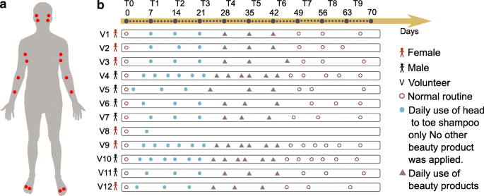
Study design and representation of changes in personal care regime over the course of 9 weeks. a Six males and six females were recruited and sampled using swabs on two locations from each body part (face, armpits, front forearms, and between toes) on the right and left side. The locations sampled were the face—upper cheek bone and lower jaw, armpit—upper and lower area, arm—front of elbow (antecubitis) and forearm (antebrachium), and feet—in between the first and second toe and third and fourth toe. Volunteers were asked to follow specific instructions for the use of skin care products. b Following the use of their personal skin care products (brown circles), all volunteers used only the same head to toe shampoo during the first 3 weeks (week 1–week 3) and no other beauty product was applied (solid blue circle). The following 3 weeks (week 4–week 6), four selected commercial beauty products were applied daily by all volunteers on the specific body part (deodorant antiperspirant for the armpits, soothing foot powder for the feet between toes, sunscreen for the face, and moisturizer for the front forearm) (triangles) and continued to use the same shampoo. During the last 3 weeks (week 7–week 9), all volunteers went back to their normal routine and used their personal beauty products (circles). Samples were collected once a week (from day 0 to day 68—10 timepoints from T0 to T9) for volunteers 1, 2, 3, 4, 5, 6, 7, 9, 10, 11, and 12, and on day 0 and day 6 for volunteer 8, who withdraw from the study after day 6. For 3 individuals (volunteers 4, 9, 10), samples were collected twice a week (19 timepoints total). Samples collected for 11 volunteers during 10 timepoints: 11 volunteers × 10 timepoints × 4 samples × 4 body sites = 1760. Samples collected from 3 selected volunteers during 9 additional timepoints: 3 volunteers × 9 timepoints × 4 samples × 4 body sites = 432. See also the “ Subject recruitment and sample collection ” section in the “ Methods ” section

Monitoring the persistence of personal care product ingredients in the armpits over a 9-week period. a Heatmap representation of the most abundant molecular features detected in the armpits of all individuals during the four phases (0: initial, 1–3: no beauty products, 4–6: common products, and 7–9: personal products). Green color in the heatmap represents the highest molecular abundance and blue color the lowest one. Orange boxes with plain lines represent enlargement of cluster of molecules that persist on the armpits of volunteer 1 ( b ) and volunteer 3 ( c , d ). Orange clusters with dotted lines represent same clusters of molecules found on the armpits of other volunteers. Orange arrows represent the cluster of compounds characteristic of the antiperspirant used during T4–T6. b Polyethylene glycol (PEG) molecular clusters that persist on the armpits of individual 1. The molecular subnetwork, representing molecular families [ 24 ], is part of a molecular network ( http://gnps.ucsd.edu/ProteoSAFe/status.jsp?task=f5325c3b278a46b29e8860ec5791d5ad ) generated from MS/MS data collected from the armpits of volunteer 1 (T0–T3) MSV000081582 and MS/MS data collected from the deodorant used by volunteer 1 before the study started (T0) MSV000081580. c , d Polypropylene glycol (PPG) molecular families that persist on the armpits of individual 3, along with the corresponding molecular subnetwork that is part of the molecular network accessible here http://gnps.ucsd.edu/ProteoSAFe/status.jsp?task=aaa1af68099d4c1a87e9a09f398fe253 . Subnetworks were generated from MS/MS data collected from the armpits of volunteer 3 (T0–T3) MSV000081582 and MS/MS data collected from the deodorant used by volunteer 3 at T0 MSV000081580. The network nodes were annotated with colors. Nodes represent MS/MS spectra found in armpit samples of individual 1 collected during T0, T1, T2, and T3 and in personal deodorant used by individual 1 (orange nodes); armpit samples of individual 1 collected during T0, T2, and T3 and personal deodorant used by individual 1 (green nodes); armpit samples of individual 3 collected during T0, T1, T2, and T3 and in personal deodorant used by individual 3 (red nodes); armpit samples of individual 3 collected during T0 and in personal deodorant used by individual 3 (blue nodes); and armpit samples of individual 3 collected during T0 and T2 and in personal deodorant used by individual 3 (purple nodes). Gray nodes represent everything else. Error bars represent standard error of the mean calculated at each timepoint from four armpit samples collected from the right and left side of each individual separately. See also Additional file 1 : Figure S1
To understand how long beauty products persist on the skin, we monitored compounds found in deodorants used by two volunteers—female 1 and female 3—before the study (T0), over the first 3 weeks (T1–T3) (Fig. 1 b). During this phase, all participants used exclusively the same body wash during showering, making it easier to track ingredients of their personal care products. The data in the first 3 weeks (T1–T3) revealed that many ingredients of deodorants used on armpits (Fig. 2 a) persist on the skin during this time and were still detected during the first 3 weeks or at least during the first week following the last day of use. Each of the compounds detected in the armpits of individuals exhibited its own unique half-life. For example, the polyethylene glycol (PEG)-derived compounds m/z 344.227, rt 143 s (Fig. 2 b, S1J); m/z 432.279, rt 158 s (Fig. 2 b, S1K); and m/z 388.253, rt 151 s (Fig. 2 b, S1L) detected on armpits of volunteer 1 have a calculated half-life of 0.5 weeks (Additional file 1 : Figure S1J-L, all p values < 1.81e−07), while polypropylene glycol (PPG)-derived molecules m/z 481.87, rt 501 s (Fig. 2 c, S1M); m/z 560.420, rt 538 s (Fig. 2 c, S1N); m/z 788.608, rt 459 s (Fig. 2 d, S1O); m/z 846.650, rt 473 s (Fig. 2 d, S1P); and m/z 444.338, rt 486 s (Fig. 2 d, S1Q) found on armpits of volunteers 3 and 1 (Fig. 2 a) have a calculated half-life ranging from 0.7 to 1.9 weeks (Additional file 1 : Figure S1M-Q, all p values < 0.02), even though they originate from the same deodorant used by each individual. For some ingredients of deodorant used by volunteer 3 on time 0 (Additional file 1 : Figure S1M, N), a decline was observed during the first week, then little to no traces of these ingredients were detected during weeks 4–6 (T4–T6), then finally these ingredients reappear again during the last 3 weeks of personal product use (T7–T9). This suggests that these ingredients are present exclusively in the personal deodorant used by volunteer 3 before the study. Because a similar deodorant (Additional file 1 : Figure S1O-Q) and a face lotion (Additional file 1 : Figure S1R) was used by volunteer 3 and volunteer 2, respectively, prior to the study, there was no decline or absence of their ingredients during weeks 4–6 (T4–T6).
Polyethylene glycol compounds (Additional file 1 : Figure S1J-L) wash out faster from the skin than polypropylene glycol (Additional file 1 : Figure S1M-Q)(HL ~ 0.5 weeks vs ~ 1.9 weeks) and faster than fatty acids used in lotions (HL ~ 1.2 weeks) (Additional file 1 : Figure S1R), consistent with their hydrophilic (PEG) and hydrophobic properties (PPG and fatty acids) [ 25 , 26 ]. This difference in hydrophobicity is also reflected in the retention time as detected by mass spectrometry. Following the linear decrease of two PPG compounds from T0 to T1, they accumulated noticeably during weeks 2 and 3 (Additional file 1 : Figure S1M, N). This accumulation might be due to other sources of PPG such as the body wash used during this period or the clothes worn by person 3. Although PPG compounds were not listed in the ingredient list of the shampoo, we manually inspected the LC-MS data collected from this product and confirmed the absence of PPG compounds in the shampoo. The data suggest that this trend is characteristic of accumulation of PPG from additional sources. These could be clothes, beds, or sheets, in agreement with the observation of these molecules found in human habitats [ 27 ] but also in the public GNPS mass spectrometry dataset MSV000079274 that investigated the chemicals from dust collected from 1053 mattresses of children.
Temporal molecular and bacterial diversity in response to personal care use
To assess the effect of discontinuing and resuming the use of skin care products on molecular and microbiota dynamics, we first evaluated their temporal diversity. Skin sites varied markedly in their initial level (T0) of molecular and bacterial diversity, with higher molecular diversity at all sites for female participants compared to males (Fig. 3 a, b, Wilcoxon rank-sum-WR test, p values ranging from 0.01 to 0.0001, from foot to arm) and higher bacterial diversity in face (WR test, p = 0.0009) and armpits (WR test, p = 0.002) for females (Fig. 3 c, d). Temporal diversity was similar across the right and left sides of each body site of all individuals (WR test, molecular diversity: all p values > 0.05; bacterial diversity: all p values > 0.20). The data show that refraining from using beauty products (T1–T3) leads to a significant decrease in molecular diversity at all sites (Fig. 3 a, b, WR test, face: p = 8.29e−07, arm: p = 7.08e−09, armpit: p = 1.13e−05, foot: p = 0.002) and bacterial diversity mainly in armpits (WR test, p = 0.03) and feet (WR test, p = 0.04) (Fig. 3 c, d). While molecular diversity declined (Fig. 3 a, b) for arms and face, bacterial diversity (Fig. 3 c, d) was less affected in the face and arms when participants did not use skin care products (T1–T3). The molecular diversity remained stable in the arms and face of female participants during common beauty products use (T4–T6) to immediately increase as soon as the volunteers went back to their normal routines (T7–T9) (WR test, p = 0.006 for the arms and face)(Fig. 3 a, b). A higher molecular (Additional file 1 : Figure S2A) and community (Additional file 1 : Figure S2B) diversity was observed for armpits and feet of all individuals during the use of antiperspirant and foot powder (T4–T6) (WR test, molecular diversity: armpit p = 8.9e−33, foot p = 1.03e−11; bacterial diversity: armpit p = 2.14e−28, foot p = 1.26e−11), followed by a molecular and bacterial diversity decrease in the armpits when their regular personal beauty product use was resumed (T7–T9) (bacterial diversity: WR test, p = 4.780e−21, molecular diversity: WR test, p = 2.159e−21). Overall, our data show that refraining from using beauty products leads to lower molecular and bacterial diversity, while resuming the use increases their diversity. Distinct variations between male and female molecular and community richness were perceived at distinct body parts (Fig. 3 a–d). Although the chemical diversity of personal beauty products does not explain these variations (Additional file 1 : Figure S2C), differences observed between males and females may be attributed to many environmental and lifestyle factors including different original skin care and different frequency of use of beauty products (Additional file 2 : Table S1), washing routines, and diet.
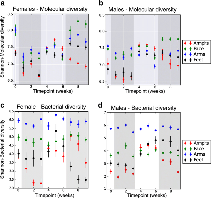
Molecular and bacterial diversity over a 9-week period, comparing samples based on their molecular (UPLC-Q-TOF-MS) or bacterial (16S rRNA amplicon) profiles. Molecular and bacterial diversity using the Shannon index was calculated from samples collected from each body part at each timepoint, separately for female ( n = 5) and male ( n = 6) individuals. Error bars represent standard error of the mean calculated at each timepoint, from up to four samples collected from the right and left side of each body part, of females ( n = 5) and males ( n = 6) separately. a , b Molecular alpha diversity measured using the Shannon index from five females (left panel) and six males (right panel), over 9 weeks, from four distinct body parts (armpits, face, arms, feet). c , d Bacterial alpha diversity measured using the Shannon index, from skin samples collected from five female (left panel) and six male individuals (right panel), over 9 weeks, from four distinct body parts (armpits, face, arms, feet). See also Additional file 1 : Figure S2
Longitudinal variation of skin metabolomics signatures
To gain insights into temporal metabolomics variation associated with beauty product use, chemical inventories collected over 9 weeks were subjected to multivariate analysis using the widely used Bray–Curtis dissimilarity metric (Fig. 4 a–c, S3A). Throughout the 9-week period, distinct molecular signatures were associated to each specific body site: arm, armpit, face, and foot (Additional file 1 : Figure S3A, Adonis test, p < 0.001, R 2 0.12391). Mass spectrometric signatures displayed distinct individual trends at each specific body site (arm, armpit, face, and foot) over time, supported by their distinct locations in PCoA (principal coordinate analysis) space (Fig. 4 a, b) and based on the Bray–Curtis distances between molecular profiles (Additional file 1 : Figure S3B, WR test, all p values < 0.0001 from T0 through T9). This suggests a high molecular inter-individual variability over time despite similar changes in personal care routines. Significant differences in molecular patterns associated to ceasing (T1–T3) (Fig. 4 b, Additional file 1 : Figure S3C, WR test, T0 vs T1–T3 p < 0.001) and resuming the use of common beauty products (T4–T6) (Additional file 1 : Figure S3C) were observed in the arm, face, and foot (Fig. 4 b), although the armpit exhibited the most pronounced changes (Fig. 4 b, Additional file 1 : Figure S3D, E, random forest highlighting that 100% of samples from each phase were correctly predicted). Therefore, we focused our analysis on this region. Molecular changes were noticeable starting the first week (T1) of discontinuing beauty product use. As shown for armpits in Fig. 4 c, these changes at the chemical level are specific to each individual, possibly due to the extremely personalized lifestyles before the study and match their original use of deodorant. Based on the initial use of underarm products (T0) (Additional file 2 : Table S1), two groups of participants can be distinguished: a group of five volunteers who used stick deodorant as evidenced by the mass spectrometry data and another group of volunteers where we found few or no traces suggesting they never or infrequently used stick deodorants (Additional file 2 : Table S1). Based on this criterion, the chemical trends shown in Fig. 4 c highlight that individuals who used stick deodorant before the beginning of the study (volunteers 1, 2, 3, 9, and 12) displayed a more pronounced shift in their armpits’ chemistries as soon as they stopped using deodorant (T1–T3), compared to individuals who had low detectable levels of stick deodorant use (volunteers 4, 6, 7, and 10), or “rarely-to-never” (volunteers 5 and 11) use stick deodorants as confirmed by the volunteers (Additional file 1 : Figure S3F, WR test, T0 vs T1–T3 all p values < 0.0001, with greater distance for the group of volunteers 1, 2, 3, 9, and 12, compared to volunteers 4, 5, 6, 7, 10, and 11). The most drastic shift in chemical profiles was observed during the transition period, when all participants applied the common antiperspirant on a daily basis (T4–T6) (Additional file 1 : Figure S3D, E). Finally, the molecular profiles became gradually more similar to those collected before the experiment (T0) as soon as the participants resumed using their personal beauty products (T7–T9) (Additional file 1 : Figure S3C), although traces of skin care products did last through the entire T7–T9 period in people who do not routinely apply these products (Fig. 4 c).

Individualized influence of beauty product application on skin metabolomics profiles over time. a Multivariate statistical analysis (principal coordinate analysis (PCoA)) comparing mass spectrometry data collected over 9 weeks from the skin of 11 individuals, all body parts, combined (first plot from the left) and then displayed separately (arm, armpits, face, feet). Color scale represents volunteer ID. The PCoA was calculated on all samples together, and subsets of the data are shown in this shared space and the other panels. b The molecular profiles collected over 9 weeks from all body parts, combined then separately (arm, armpits, face, feet). c Representative molecular profiles collected over 9 weeks from armpits of 11 individuals (volunteers 1, 2, 3, 4, 5, 6, 7, 9, 10, 11, 12). Color gradient in b and c represents timepoints (time 0 to time 9), ranging from the lightest orange color to the darkest one that represent the earliest (time 0) to the latest (time 9) timepoint, respectively. 0.5 timepoints represent additional timepoints where three selected volunteers were samples (volunteers 4, 9, and 10). PCoA plots were generated using the Bray–Curtis dissimilarity matrix and visualized in Emperor [ 28 ]. See also Additional file 1 : Figure S3
Comparing chemistries detected in armpits at the end timepoints—when no products were used (T3) and during product use (T6)—revealed distinct molecular signatures characteristic of each phase (random forest highlighting that 100% of samples from each group were correctly predicted, see Additional file 1 : Figure S3D, E). Because volunteers used the same antiperspirant during T4–T6, molecular profiles converged during that time despite individual patterns at T3 (Fig. 4 b, c, Additional file 1 : Figure S3D). These distinct chemical patterns reflect the significant impact of beauty products on skin molecular composition. Although these differences may in part be driven by beauty product ingredients detected on the skin (Additional file 1 : Figure S1), we anticipated that additional host- and microbe-derived molecules may also be involved in these molecular changes.
To characterize the chemistries that vary over time, we used molecular networking, a MS visualization approach that evaluates the relationship between MS/MS spectra and compares them to reference MS/MS spectral libraries of known compounds [ 29 , 30 ]. We recently showed that molecular networking can successfully organize large-scale mass spectrometry data collected from the human skin surface [ 18 , 19 ]. Briefly, molecular networking uses the MScluster algorithm [ 31 ] to merge all identical spectra and then compares and aligns all unique pairs of MS/MS spectra based on their similarities where 1.0 indicates a perfect match. Similarities between MS/MS spectra are calculated using a similarity score, and are interpreted as molecular families [ 19 , 24 , 32 , 33 , 34 ]. Here, we used this method to compare and characterize chemistries found in armpits, arms, face, and foot of 11 participants. Based on MS/MS spectral similarities, chemistries highlighted through molecular networking (Additional file 1 : Figure S4A) were associated with each body region with 8% of spectra found exclusively in the arms, 12% in the face, 14% in the armpits, and 2% in the foot, while 18% of the nodes were shared between all four body parts and the rest of spectra were shared between two body sites or more (Additional file 1 : Figure S4B). Greater spectral similarities were highlighted between armpits, face, and arm (12%) followed by the arm and face (9%) (Additional file 1 : Figure S4B).
Molecules were annotated with Global Natural Products Social Molecular Networking (GNPS) libraries [ 29 ], using accurate parent mass and MS/MS fragmentation patterns, according to level 2 or 3 of annotation defined by the 2007 metabolomics standards initiative [ 35 ]. Through annotations, molecular networking revealed that many compounds derived from steroids (Fig. 5 a–d), bile acids (Additional file 1 : Figure S5A-D), and acylcarnitines (Additional file 1 : Figure S5E-F) were exclusively detected in the armpits. Using authentic standards, the identity of some pheromones and bile acids were validated to a level 1 identification with matched retention times (Additional file 1 : Figure S6B, S7A, C, D). Other steroids and bile acids were either annotated using standards with identical MS/MS spectra but slightly different retention times (Additional file 1 : Figure S6A) or annotated with MS/MS spectra match with reference MS/MS library spectra (Additional file 1 : Figure S6C, D, S7B, S6E-G). These compounds were therefore classified as level 3 [ 35 ]. Acylcarnitines were annotated to a family of possible acylcarnitines (we therefore classify as level 3), as the positions of double bonds or cis vs trans configurations are unknown (Additional file 1 : Figure S8A, B).
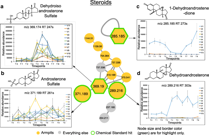
Underarm steroids and their longitudinal abundance. a – d Steroid molecular families in the armpits and their relative abundance over a 9-week period. Molecular networking was applied to characterize chemistries from the skin of 11 healthy individuals. The full network is shown in Additional file 1 : Figure S4A, and networking parameters can be found here http://gnps.ucsd.edu/ProteoSAFe/status.jsp?task=284fc383e4c44c4db48912f01905f9c5 for MS/MS datasets MSV000081582. Each node represents a consensus of a minimum of 3 identical MS/MS spectra. Yellow nodes represent MS/MS spectra detected in armpits samples. Hexagonal shape represents MS/MS spectra match between skin samples and chemical standards. Plots are representative of the relative abundance of each compound over time, calculated separately from LC-MS1 data collected from the armpits of each individual. Steroids detected in armpits are a , dehydroisoandrosterone sulfate ( m/z 369.190, rt 247 s), b androsterone sulfate ( m/z 371.189, rt 261 s), c 1-dehydroandrostenedione ( m/z 285.185, rt 273 s), and d dehydroandrosterone ( m/z 289.216, rt 303 s). Relative abundance over time of each steroid compound is represented. Error bars represent the standard error of the mean calculated at each timepoint from four armpit samples from the right and left side of each individual separately. See also Additional file 1 : Figures S4-S8
Among the steroid compounds, several molecular families were characterized: androsterone (Fig. 5 a, b, d), androstadienedione (Fig. 5 c), androstanedione (Additional file 1 : Figure S6E), androstanolone (Additional file 1 : Figure S6F), and androstenedione (Additional file 1 : Figure S6G). While some steroids were detected in the armpits of several individuals, such as dehydroisoandrosterone sulfate ( m/z 369.19, rt 247 s) (9 individuals) (Fig. 5 a, Additional file 1 : Figure S6A), androsterone sulfate ( m/z 371.189, rt 261 s) (9 individuals) (Fig. 5 b, Additional file 1 : Figure S6C), and 5-alpha-androstane-3,17-dione ( m/z 271.205, rt 249 s) (9 individuals) (Additional file 1 : Figure S6E), other steroids including 1-dehydroandrostenedione ( m/z 285.185, rt 273 s) (Fig. 5 c, Additional file 1 : Figure S6B), dehydroandrosterone ( m/z 289.216, rt 303 s) (Fig. 5 d, Additional file 1 : Figure S6D), and 5-alpha-androstan-17.beta-ol-3-one ( m/z 291.231, rt 318 s) (Additional file 1 : Figure S6F) were only found in the armpits of volunteer 11 and 4-androstene-3,17-dione ( m/z 287.200, rt 293 s) in the armpits of volunteer 11 and volunteer 5, both are male that never applied stick deodorants (Additional file 1 : Figure S6G). Each molecular species exhibited a unique pattern over the 9-week period. The abundance of dehydroisoandrosterone sulfate (Fig. 5 a, WR test, p < 0.01 for 7 individuals) and dehydroandrosterone (Fig. 5 a, WR test, p = 0.00025) significantly increased during the use of antiperspirant (T4–T6), while androsterone sulfate (Fig. 5 b) and 5-alpha-androstane-3,17-dione (Additional file 1 : Figure S6E) display little variation over time. Unlike dehydroisoandrosterone sulfate (Fig. 5 a) and dehydroandrosterone (Fig. 5 d), steroids including 1-dehydroandrostenedione (Fig. 5 c, WR test, p = 0.00024) and 4-androstene-3,17-dione (Additional file 1 : Figure S6G, WR test, p = 0.00012) decreased in abundance during the 3 weeks of antiperspirant application (T4–T6) in armpits of male 11, and their abundance increased again when resuming the use of his normal skin care routines (T7–T9). Interestingly, even within the same individual 11, steroids were differently impacted by antiperspirant use as seen for 1-dehydroandrostenedione that decreased in abundance during T4–T6 (Fig. 5 c, WR test, p = 0.00024), while dehydroandrosterone increased in abundance (Fig. 5 d, WR test, p = 0.00025), and this increase was maintained during the last 3 weeks of the study (T7–T9).
In addition to steroids, many bile acids (Additional file 1 : Figure S5A-D) and acylcarnitines (Additional file 1 : Figure S5E-F) were detected on the skin of several individuals through the 9-week period. Unlike taurocholic acid found only on the face (Additional file 1 : Figures S5A, S7A) and tauroursodeoxycholic acid detected in both armpits and arm samples (Additional file 1 : Figures S5B, S7B), other primary bile acids such as glycocholic (Additional file 1 : Figures S5C, S7C) and chenodeoxyglycocholic acid (Additional file 1 : Figures S5D, S7D) were exclusively detected in the armpits. Similarly, acylcarnitines were also found either exclusively in the armpits (hexadecanoyl carnitines) (Additional file 1 : Figures S5E, S8A) or in the armpits and face (tetradecenoyl carnitine) (Additional file 1 : Figures S5F, S8B) and, just like the bile acids, they were also stably detected during the whole 9-week period.
Bacterial communities and their variation over time
Having demonstrated the impact of beauty products on the chemical makeup of the skin, we next tested the extent to which skin microbes are affected by personal care products. We assessed temporal variation of bacterial communities detected on the skin of healthy individuals by evaluating dissimilarities of bacterial collections over time using unweighted UniFrac distance [ 36 ] and community variation at each body site in association to beauty product use [ 3 , 15 , 37 ]. Unweighted metrics are used for beta diversity calculations because we are primarily concerned with changes in community membership rather than relative abundance. The reason for this is that skin microbiomes can fluctuate dramatically in relative abundance on shorter timescales than that assessed here. Longitudinal variations were revealed for the armpits (Fig. 6 a) and feet microbiome by their overall trend in the PCoA plots (Fig. 6 b), while the arm (Fig. 6 c) and face (Fig. 6 d) displayed relatively stable bacterial profiles over time. As shown in Fig. 6 a–d, although the microbiome was site-specific, it varied more between individuals and this inter-individual variability was maintained over time despite same changes in personal care routine (WR test, all p values at all timepoints < 0.05, T5 p = 0.07), in agreement with previous findings that individual differences in the microbiome are large and stable over time [ 3 , 4 , 10 , 37 ]. However, we show that shifts in the microbiome can be induced by changing hygiene routine and therefore skin chemistry. Changes associated with using beauty products (T4–T6) were more pronounced for the armpits (Fig. 6 a, WR test, p = 1.61e−52) and feet (Fig. 6 b, WR test, p = 6.15e−09), while little variations were observed for the face (Fig. 6 d, WR test, p = 1.402.e−83) and none for the arms (Fig. 6 c, WR test, p = 0.296).
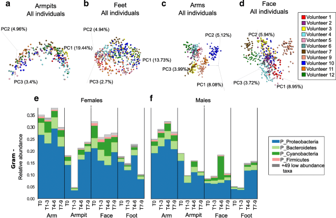
Longitudinal variation of skin bacterial communities in association with beauty product use. a - d Bacterial profiles collected from skin samples of 11 individuals, over 9 weeks, from four distinct body parts a) armpits, b) feet, c) arms and d) face, using multivariate statistical analysis (Principal Coordinates Analysis PCoA) and unweighted Unifrac metric. Each color represents bacterial samples collected from an individual. PCoA were calculated separately for each body part. e , f Representative Gram-negative (Gram -) bacteria collected from arms, armpits, face and feet of e) female and f) male participants. See also Additional file 1 : Figure S9A, B showing Gram-negative bacterial communities represented at the genus level
A significant increase in abundance of Gram-negative bacteria including the phyla Proteobacteria and Bacteroidetes was noticeable for the armpits and feet of both females (Fig. 6 e; Mann–Whitney U , p = 8.458e−07) and males (Fig. 6 f; Mann–Whitney U , p = 0.0004) during the use of antiperspirant (T4–T6), while their abundance remained stable for the arms and face during that time (Fig. 6 e, f; female arm p = 0.231; female face p value = 0.475; male arm p = 0.523;male face p = 6.848751e−07). These Gram-negative bacteria include Acinetobacter and Paracoccus genera that increased in abundance in both armpits and feet of females (Additional file 1 : Figure S9A), while a decrease in abundance of Enhydrobacter was observed in the armpits of males (Additional file 1 : Figure S9B). Cyanobacteria, potentially originating from plant material (Additional file 1 : Figure S9C) also increased during beauty product use (T4–T6) especially in males, in the armpits and face of females (Fig. 6 e) and males (Fig. 6 f). Interestingly, although chloroplast sequences (which group phylogenetically within the cyanobacteria [ 38 ]) were only found in the facial cream (Additional file 1 : Figure S9D), they were detected in other locations as well (Fig. 6 e, f. S9E, F), highlighting that the application of a product in one region will likely affect other regions of the body. For example, when showering, a face lotion will drip down along the body and may be detected on the feet. Indeed, not only did the plant material from the cream reveal this but also the shampoo used for the study for which molecular signatures were readily detected on the feet as well (Additional file 1 : Figure S10A). Minimal average changes were observed for Gram-positive organisms (Additional file 1 : Figure S10B, C), although in some individuals the variation was greater than others (Additional file 1 : Figure S10D, E) as discussed for specific Gram-positive taxa below.
At T0, the armpit’s microflora was dominated by Staphylococcus (26.24%, 25.11% of sequencing reads for females and 27.36% for males) and Corynebacterium genera (26.06%, 17.89% for females and 34.22% for males) (Fig. 7 a—first plot from left and Additional file 1 : Figure S10D, E). They are generally known as the dominant armpit microbiota and make up to 80% of the armpit microbiome [ 39 , 40 ]. When no deodorants were used (T1–T3), an overall increase in relative abundance of Staphylococcus (37.71%, 46.78% for females and 30.47% for males) and Corynebacterium (31.88%, 16.50% for females and 44.15% for males) genera was noticeable (WR test, p < 3.071e−05) (Fig. 7 a—first plot from left), while the genera Anaerococcus and Peptoniphilus decreased in relative abundance (WR test, p < 0.03644) (Fig. 7 a—first plot from left and Additional file 1 : Figure S10D, E). When volunteers started using antiperspirants (T4–T6), the relative abundance of Staphylococcus (37.71%, 46.78% females and 30.47% males, to 21.71%, 25.02% females and 19.25% males) and Corynebacterium (31.88%, 16.50% females and 44.15% males, to 15.83%, 10.76% females and 19.60% males) decreased (WR test, p < 3.071e−05) (Fig. 7 a, Additional file 1 : Figure S10D, E) and at the same time, the overall alpha diversity increased significantly (WR test, p = 3.47e−11) (Fig. 3 c, d). The microbiota Anaerococcus (WR test, p = 0.0006018) , Peptoniphilus (WR test, p = 0.008639), and Micrococcus (WR test, p = 0.0377) increased significantly in relative abundance, together with a lot of additional low-abundant species that lead to an increase in Shannon alpha diversity (Fig. 3 c, d). When participants went back to normal personal care products (T7–T9), the underarm microbiome resembled the original underarm community of T0 (WR test, p = 0.7274) (Fig. 7 a). Because armpit bacterial communities are person-specific (inter-individual variability: WR test, all p values at all timepoints < 0.05, besides T5 p n.s), variation in bacterial abundance upon antiperspirant use (T4–T6) differ between individuals and during the whole 9-week period (Fig. 7a —taxonomic plots per individual). For example, the underarm microbiome of male 5 exhibited a unique pattern, where Corynebacterium abundance decreased drastically during the use of antiperspirant (82.74 to 11.71%, WR test, p = 3.518e−05) while in the armpits of female 9 a huge decrease in Staphylococcus abundance was observed (Fig. 7 a) (65.19 to 14.85%, WR test, p = 0.000113). Unlike other participants, during T0–T3, the armpits of individual 11 were uniquely characterized by the dominance of a sequence that matched most closely to the Enhydrobacter genera . The transition to antiperspirant use (T4–T6) induces the absence of Enhydrobacter (30.77 to 0.48%, WR test, p = 0.01528) along with an increase of Corynebacterium abundance (26.87 to 49.74%, WR test, p = 0.1123) (Fig. 7 a—male 11).
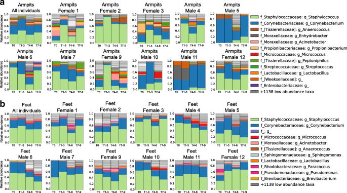
Person-to-person bacterial variabilities over time in the armpits and feet. a Armpit microbiome changes when stopping personal care product use, then resuming. Armpit bacterial composition of the 11 volunteers combined, then separately, (female 1, female 2, female 3, male 4, male 5, male 6, male 7, female 9, male 10, male 11, female 12) according to the four periods within the experiment. b Feet bacterial variation over time of the 12 volunteers combined, then separately (female 1, female 2, female 3, male 4, male 5, male 6, male 7, female 9, male 10, male 11, female 12) according to the four periods within the experiment. See also Additional file 1 : Figure S9-S13
In addition to the armpits, a decline in abundance of Staphylococcus and Corynebacterium was perceived during the use of the foot powder (46.93% and 17.36%, respectively) compared to when no beauty product was used (58.35% and 22.99%, respectively) (WR test, p = 9.653e−06 and p = 0.02032, respectively), while the abundance of low-abundant foot bacteria significantly increased such as Micrococcus (WR test, p = 1.552e−08), Anaerococcus (WR test, p = 3.522e−13), Streptococcus (WR test, p = 1.463e−06), Brevibacterium (WR test, p = 6.561e−05), Moraxellaceae (WR test, p = 0.0006719), and Acinetobacter (WR test, p = 0.001487), leading to a greater bacterial diversity compared to other phases of the study (Fig. 7 b first plot from left, Additional file 1 : Figure S10D, E, Fig. 3 c, d).
We further evaluated the relationship between the two omics datasets by superimposing the principal coordinates calculated from metabolome and microbiome data (Procrustes analysis) (Additional file 1 : Figure S11) [ 34 , 41 , 42 ]. Metabolomics data were more correlated with patterns observed in microbiome data in individual 3 (Additional file 1 : Figure S11C, Mantel test, r = 0.23, p < 0.001), individual 5 (Additional file 1 : Figure S11E, r = 0.42, p < 0.001), individual 9 (Additional file 1 : Figure S11H, r = 0.24, p < 0.001), individual 10 (Additional file 1 : Figure S11I, r = 0.38, p < 0.001), and individual 11 (Additional file 1 : Figure S11J, r = 0.35, p < 0.001) when compared to other individuals 1, 2, 4, 6, 7, and 12 (Additional file 1 : Figure S11A, B, D, F, G, K, respectively) (Mantel test, all r < 0.2, all p values < 0.002, for volunteer 2 p n.s). Furthermore, these correlations were individually affected by ceasing (T1–T3) or resuming the use of beauty products (T4–T6 and T7–T9) (Additional file 1 : Figure S11A-K).
Overall, metabolomics–microbiome correlations were consistent over time for the arms, face, and feet although alterations were observed in the arms of volunteers 7 (Additional file 1 : Figure S11G) and 10 (Additional file 1 : Figure S11I) and the face of volunteer 7 (Additional file 1 : Figure S11G) during product use (T4–T6). Molecular–bacterial correlations were mostly affected in the armpits during antiperspirant use (T4–T6), as seen for volunteers male 7 (Additional file 1 : Figure S11G) and 11 (Additional file 1 : Figure S11J) and females 2 (Additional file 1 : Figure S11B), 9 (Additional file 1 : Figure S11H), and 12 (Additional file 1 : Figure S11K). This perturbation either persisted during the last 3 weeks (Additional file 1 : Figure S11D, E, H, I, K) when individuals went back to their normal routine (T7–T9) or resembled the initial molecular–microbial correlation observed in T0 (Additional file 1 : Figure S11C, G, J). These alterations in molecular–bacterial correlation are driven by metabolomics changes during antiperspirant use as revealed by metabolomics shifts on the PCoA space (Additional file 1 : Figure S11), partially due to the deodorant’s chemicals (Additional file 1 : Figure S1J, K) but also to changes observed in steroid levels in the armpits (Fig. 5A, C, D , Additional file 1 : Figure S6G), suggesting metabolome-dependant changes of the skin microbiome. In agreement with previous findings that showed efficient biotransformation of steroids by Corynebacterium [ 43 , 44 ], our correlation analysis associates specific steroids that were affected by antiperspirant use in the armpits of volunteer 11 (Fig. 5 c, d, Additional file 1 : Figure S6G) with microbes that may produce or process them: 1-dehydroandrostenedione, androstenedione, and dehydrosterone with Corynebacterium ( r = − 0.674, p = 6e−05; r = 0.671, p = 7e−05; r = 0.834, p < 1e−05, respectively) (Additional file 1 : Figure S12A, B, C, respectively) and Enhydrobacter ( r = 0.683, p = 4e−05; r = 0.581, p = 0.00095; r = 0.755, p < 1e−05 respectively) (Additional file 1 : Figure S12D, E, F, respectively).
Despite the widespread use of skin care and hygiene products, their impact on the molecular and microbial composition of the skin is poorly studied. We established a workflow that examines individuals to systematically study the impact of such lifestyle characteristics on the skin by taking a broad look at temporal molecular and bacterial inventories and linking them to personal skin care product use. Our study reveals that when the hygiene routine is modified, the skin metabolome and microbiome can be altered, but that this alteration depends on product use and location on the body. We also show that like gut microbiome responses to dietary changes [ 20 , 21 ], the responses are individual-specific.
We recently reported that traces of our lifestyle molecules can be detected on the skin days and months after the original application [ 18 , 19 ]. Here, we show that many of the molecules associated with our personal skin and hygiene products had a half-life of 0.5 to 1.9 weeks even though the volunteers regularly showered, swam, or spent time in the ocean. Thus, a single application of some of these products has the potential to alter the microbiome and skin chemistry for extensive periods of time. Our data suggests that although host genetics and diet may play a role, a significant part of the resilience of the microbiome that has been reported [ 10 , 45 ] is due to the resilience of the skin chemistry associated with personal skin and hygiene routines, or perhaps even continuous re-exposure to chemicals from our personal care routines that are found on mattresses, furniture, and other personal objects [ 19 , 27 , 46 ] that are in constant contact. Consistent with this observation is that individuals in tribal regions and remote villages that are infrequently exposed to the types of products used in this study have very different skin microbial communities [ 47 , 48 ] and that the individuals in this study who rarely apply personal care products had a different starting metabolome. We observed that both the microbiome and skin chemistry of these individuals were most significantly affected by these products. This effect by the use of products at T4–T6 on the volunteers that infrequently used them lasted to the end phase of the study even though they went back to infrequent use of personal care products. What was notable and opposite to what the authors originally hypothesized is that the use of the foot powder and antiperspirant increased the diversity of microbes and that some of this diversity continued in the T7–T9 phase when people went back to their normal skin and hygiene routines. It is likely that this is due to the alteration in the nutrient availability such as fatty acids and moisture requirements, or alteration of microbes that control the colonization via secreted small molecules, including antibiotics made by microbes commonly found on the skin [ 49 , 50 ].
We detected specific molecules on the skin that originated from personal care products or from the host. One ingredient that lasts on the skin is propylene glycol, which is commonly used in deodorants and antiperspirants and added in relatively large amounts as a humectant to create a soft and sleek consistency [ 51 ]. As shown, daily use of personal care products is leading to high levels of exposure to these polymers. Such polymers cause contact dermatitis in a subset of the population [ 51 , 52 ]. Our data reveal a lasting accumulation of these compounds on the skin, suggesting that it may be possible to reduce their dose in deodorants or frequency of application and consequently decrease the degree of exposure to such compounds. Formulation design of personal care products may be influenced by performing detailed outcome studies. In addition, longer term impact studies are needed, perhaps in multiple year follow-up studies, to assess if the changes we observed are permanent or if they will recover to the original state.
Some of the host- and microbiome-modified molecules were also detected consistently, such as acylcarnitines, bile acids, and certain steroids. This means that a portion of the molecular composition of a person’s skin is not influenced by the beauty products applied to the skin, perhaps reflecting the level of exercise for acylcarnitines [ 53 , 54 ] or the liver (dominant location where they are made) or gallbladder (where they are stored) function for bile acids. The bile acid levels are not related to sex and do not change in amount during the course of this study. While bile acids are typically associated with the human gut microbiome [ 34 , 55 , 56 , 57 , 58 ], it is unclear what their role is on the skin and how they get there. One hypothesis is that they are present in the sweat that is excreted through the skin, as this is the case for several food-derived molecules such as caffeine or drugs and medications that have been previously reported on the human skin [ 19 ] or that microbes synthesize them de novo [ 55 ]. The only reports we could find on bile acids being associated with the skin describe cholestasis and pruritus diseases. Cholestasis and pruritus in hepatobiliary disease have symptoms of skin bile acid accumulation that are thought to be responsible for severe skin itching [ 59 , 60 ]. However, since bile acids were found in over 50% of the healthy volunteers, their detection on the skin is likely a common phenotype among the general population and not only reflective of disease, consistent with recent reports challenging these molecules as biomarkers of disease [ 59 ]. Other molecules that were detected consistently came from personal care products.
Aside from molecules that are person-specific and those that do not vary, there are others that can be modified via personal care routines. Most striking is how the personal care routines influenced changes in hormones and pheromones in a personalized manner. This suggests that there may be personalized recipes that make it possible to make someone more or less attractive to others via adjustments of hormonal and pheromonal levels through alterations in skin care.
Here, we describe the utilization of an approach that combines metabolomics and microbiome analysis to assess the effect of modifying personal care regime on skin chemistry and microbes. The key findings are as follows: (1) Compounds from beauty products last on the skin for weeks after their first use despite daily showering. (2) Beauty products alter molecular and bacterial diversity as well as the dynamic and structure of molecules and bacteria on the skin. (3) Molecular and bacterial temporal variability is product-, site-, and person-specific, and changes are observed starting the first week of beauty product use. This study provides a framework for future investigations to understand how lifestyle characteristics such as diet, outdoor activities, exercise, and medications shape the molecular and microbial composition of the skin. These factors have been studied far more in their impact on the gut microbiome and chemistry than in the skin. Revealing how such factors can affect skin microbes and their associated metabolites may be essential to define long-term skin health by restoring the appropriate microbes particularly in the context of skin aging [ 61 ] and skin diseases [ 49 ] as has shown to be necessary for amphibian health [ 62 , 63 ], or perhaps even create a precision skin care approach that utilizes the proper care ingredients based on the microbial and chemical signatures that could act as key players in host defense [ 49 , 64 , 65 ].
Subject recruitment and sample collection
Twelve individuals between 25 and 40 years old were recruited to participate in this study, six females and six males. Female volunteer 8 dropped out of the study as she developed a skin irritation during the T1–T3 phase. All volunteers signed a written informed consent in accordance with the sampling procedure approved by the UCSD Institutional Review Board (Approval Number 161730). Volunteers were required to follow specific instructions during 9 weeks. They were asked to bring in samples of their personal care products they used prior to T0 so they could be sampled as well. Following the initial timepoint time 0 and during the first 3 weeks (week 1–week 3), volunteers were asked not to use any beauty products (Fig. 1 b). During the next 3 weeks (week 4–week 6), four selected commercial beauty products provided to all volunteers were applied once a day at specific body part (deodorant for the armpits, soothing foot powder between the toes, sunscreen for the face, and moisturizer for front forearms) (Fig. 1 b, Additional file 3 : Table S2 Ingredient list of beauty products). During the first 6 weeks, volunteers were asked to shower with a head to toe shampoo. During the last 3 weeks (week 7–week 9), all volunteers went back to their normal routine and used the personal care products used before the beginning of the study (Fig. 1 b). Volunteers were asked not to shower the day before sampling. Samples were collected by the same three researchers to ensure consistency in sampling and the area sampled. Researchers examined every subject together and collected metabolomics and microbiome samples from each location together. Samples were collected once a week (from day 0 to day 68—10 timepoints total) for volunteers 1, 2, 3, 4, 5, 6, 7, 9, 10, 11, and 12, and on day 0 and day 6 for volunteer 8. For individuals 4, 9, and 10, samples were collected twice a week. Samples collected for 11 volunteers during 10 timepoints: 11 volunteers × 10 timepoints × 4 samples × 4 body sites = 1760. Samples collected from 3 selected volunteers during 9 additional timepoints: 3 volunteers × 9 timepoints × 4 samples × 4 body sites = 432. All samples were collected following the same protocol described in [ 18 ]. Briefly, samples were collected over an area of 2 × 2 cm, using pre-moistened swabs in 50:50 ethanol/water solution for metabolomics analysis or in Tris-EDTA buffer for 16S rRNA sequencing. Four samples were collected from each body part right and left side. The locations sampled were the face—upper cheek bone and lower jaw, armpit—upper and lower area, arm—front of the elbow (antecubitis) and forearm (antebrachium), and feet—in between the first and second toe and third and fourth toe. Including personal care product references, a total of 2275 samples were collected over 9 weeks and were submitted to both metabolomics and microbial inventories.
Metabolite extraction and UPLC-Q-TOF mass spectrometry analysis
Skin swabs were extracted and analyzed using a previously validated workflow described in [ 18 , 19 ]. All samples were extracted in 200 μl of 50:50 ethanol/water solution for 2 h on ice then overnight at − 20 °C. Swab sample extractions were dried down in a centrifugal evaporator then resuspended by vortexing and sonication in a 100 μl 50:50 ethanol/water solution containing two internal standards (fluconazole 1 μM and amitriptyline 1 μM). The ethanol/water extracts were then analyzed using a previously validated UPLC-MS/MS method [ 18 , 19 ]. We used a ThermoScientific UltiMate 3000 UPLC system for liquid chromatography and a Maxis Q-TOF (Quadrupole-Time-of-Flight) mass spectrometer (Bruker Daltonics), controlled by the Otof Control and Hystar software packages (Bruker Daltonics) and equipped with ESI source. UPLC conditions of analysis are 1.7 μm C18 (50 × 2.1 mm) UHPLC Column (Phenomenex), column temperature 40 °C, flow rate 0.5 ml/min, mobile phase A 98% water/2% acetonitrile/0.1% formic acid ( v / v ), mobile phase B 98% acetonitrile/2% water/0.1% formic acid ( v / v ). A linear gradient was used for the chromatographic separation: 0–2 min 0–20% B, 2–8 min 20–99% B, 8–9 min 99–99% B, 9–10 min 0% B. Full-scan MS spectra ( m/z 80–2000) were acquired in a data-dependant positive ion mode. Instrument parameters were set as follows: nebulizer gas (nitrogen) pressure 2 Bar, capillary voltage 4500 V, ion source temperature 180 °C, dry gas flow 9 l/min, and spectra rate acquisition 10 spectra/s. MS/MS fragmentation of 10 most intense selected ions per spectrum was performed using ramped collision induced dissociation energy, ranged from 10 to 50 eV to get diverse fragmentation patterns. MS/MS active exclusion was set after 4 spectra and released after 30 s.
Mass spectrometry data collected from the skin of 12 individuals can be found here MSV000081582.
LC-MS data processing
LC-MS raw data files were converted to mzXML format using Compass Data analysis software (Bruker Daltonics). MS1 features were selected for all LC-MS datasets collected from the skin of 12 individuals and blank samples (total 2275) using the open-source software MZmine [ 66 ]—see Additional file 4 : Table S3 for parameters. Subsequent blank filtering, total ion current, and internal standard normalization were performed (Additional file 5 : Table S4) for representation of relative abundance of molecular features (Fig. 2 , Additional file 1 : Figure S1), principal coordinate analysis (PCoA) (Fig. 4 ). For steroid compounds in Fig. 5 a–d, bile acids (Additional file 1 : Figure S5A-D), and acylcarnitines (Additional file 1 : Figure S5E, F) compounds, crop filtering feature available in MZmine [ 66 ] was used to identify each feature separately in all LC-MS data collected from the skin of 12 individuals (see Additional file 4 : Table S3 for crop filtering parameters and feature finding in Additional file 6 : Table S5).
Heatmap in Fig. 2 was constructed from the bucket table generated from LC-MS1 features (Additional file 7 : Table S6) and associated metadata (Additional file 8 : Table S7) using the Calour command line available here: https://github.com/biocore/calour . Calour parameters were as follows: normalized read per sample 5000 and cluster feature minimum reads 50. Procrustes and Pearson correlation analyses in Additional file 1 : Figures S10 and S11 were performed using the feature table in Additional file 9 : Table S8, normalized using the probabilistic quotient normalization method [ 67 ].
16S rRNA amplicon sequencing
16S rRNA sequencing was performed following the Earth Microbiome Project protocols [ 68 , 69 ], as described before [ 18 ]. Briefly, DNA was extracted using MoBio PowerMag Soil DNA Isolation Kit and the V4 region of the 16S rRNA gene was amplified using barcoded primers [ 70 ]. PCR was performed in triplicate for each sample, and V4 paired-end sequencing [ 70 ] was performed using Illumina HiSeq (La Jolla, CA). Raw sequence reads were demultiplexed and quality controlled using the defaults, as provided by QIIME 1.9.1 [ 71 ]. The primary OTU table was generated using Qiita ( https://qiita.ucsd.edu/ ), using UCLUST ( https://academic.oup.com/bioinformatics/article/26/19/2460/230188 ) closed-reference OTU picking method against GreenGenes 13.5 database [ 72 ]. Sequences can be found in EBI under accession number EBI: ERP104625 or in Qiita ( qiita.ucsd.edu ) under Study ID 10370. Resulting OTU tables were then rarefied to 10,000 sequences/sample for downstream analyses (Additional file 10 Table S9). See Additional file 11 : Table S10 for read count per sample and Additional file 1 : Figure S13 representing the samples that fall out with rarefaction at 10,000 threshold. The dataset includes 35 blank swab controls and 699 empty controls. The blank samples can be accessed through Qiita ( qiita.ucsd.edu ) as study ID 10370 and in EBI with accession number EBI: ERP104625. Blank samples can be found under the metadata category “sample_type” with the name “empty control” and “Swabblank.” These samples fell below the rarefaction threshold at 10,000 (Additional file 11 : Table S10).
To rule out the possibility that personal care products themselves contained the microbes that induced the changes in the armpit and foot microbiomes that were observed in this study (Fig. 7 ), we subjected the common personal care products that were used in this study during T4–T6 also to 16S rRNA sequencing. The data revealed that within the limit of detectability of the current experiment, few 16S signatures were detected. One notable exception was the most dominant plant-originated bacteria chloroplast detected in the sunscreen lotion applied on the face (Additional file 1 : Figure S9D), that was also detected on the face of individuals and at a lower level on their arms, sites where stable microbial communities were observed over time (Additional file 1 : Figure S9E, F). This finding is in agreement with our previous data from the 3D cartographical skin maps that revealed the presence of co-localized chloroplast and lotion molecules [ 18 ]. Other low-abundant microbial signatures found in the sunscreen lotion include additional plant-associated bacteria: mitochondria [ 73 ], Bacillaceae [ 74 , 75 ], Planococcaceae [ 76 ], and Ruminococcaceae family [ 77 ], but all these bacteria are not responsible for microbial changes associated to beauty product use, as they were poorly detected in the armpits and feet (Fig. 7 ).
To assess the origin of Cyanobacteria detected in skin samples, each Greengenes [ 72 ] 13_8 97% OTU table (per lane; obtained from Qiita [ 78 ] study 10,370) was filtered to only features with a p__Cyanobacteria phylum. The OTU maps for these tables—which relate each raw sequence to an OTU ID—were then filtered to only those observed p__Cyanobacteria OTU IDs. The filtered OTU map was used to extract the raw sequences into a single file. Separately, the unaligned Greengenes 13_8 99% representative sequences were filtered into two sets, first the set of representatives associated with c__Chloroplast (our interest database), and second the set of sequences associated with p__Cyanobacteria without the c__Chloroplast sequences (our background database). Platypus Conquistador [ 79 ] was then used to determine what reads were observed exclusively in the interest database and not in the background database. Of the 4,926,465 raw sequences associated with a p__Cyanobacteria classification (out of 318,686,615 total sequences), at the 95% sequence identity level with 100% alignment, 4,860,258 sequences exclusively recruit to full-length chloroplast 16S by BLAST [ 80 ] with the bulk recruiting to streptophytes (with Chlorophyta and Stramenopiles to a lesser extent). These sequences do not recruit non-chloroplast Cyanobacteria full length 16S.
Half-life calculation for metabolomics data
In order to estimate the biological half-life of molecules detected in the skin, the first four timepoints of the study (T0, T1, T2, T3) were considered for the calculation to allow the monitoring of personal beauty products used at T0. The IUPAC’s definition of biological half-life as the time required to a substance in a biological system to be reduced to half of its value, assuming an approximately exponential removal [ 81 ] was used. The exponential removal can be described as C ( t ) = C 0 e − tλ where t represents the time in weeks, C 0 represents the initial concentration of the molecule, C ( t ) represents the concentration of the molecule at time t , and λ is the rate of removal [ http://onlinelibrary.wiley.com/doi/10.1002/9780470140451.ch2/summary ]. The parameter λ was estimated by a mixed linear effects model in order to account for the paired sample structure. The regression model tests the null hypothesis that λ is equal to zero and only the significant ( p value < 0.05) parameters were considered.
Principal coordinate analysis
We performed principal coordinate analysis (PCoA) on both metabolomics and microbiome data. For metabolomics, we used MS1 features (Additional file 5 : Table S4) and calculated Bray–Curtis dissimilarity metric using ClusterApp ( https://github.com/mwang87/q2_metabolomics ).
For microbiome data, we used rarefied OTU table (Additional file 10 : Table S9) and used unweighted UniFrac metric [ 36 ] to calculate beta diversity distance matrix using QIIME2 (https://qiime2.org). Results from both data sources were visualized using Emperor ( https://biocore.github.io/emperor/ ) [ 28 ].
Molecular networking
Molecular networking was generated from LC-MS/MS data collected from skin samples of 11 individuals MSV000081582, using the Global Natural Products Social Molecular Networking platform (GNPS) [ 29 ]. Molecular network parameters for MS/MS data collected from all body parts of 11 individuals during T0–T9 MSV000081582 are accessible here http://gnps.ucsd.edu/ProteoSAFe/status.jsp?task=284fc383e4c44c4db48912f01905f9c5 . Molecular network parameters for MS/MS data collected from armpits T0–T3 MSV000081582 and deodorant used by individual 1 and 3 MSV000081580 can be found here http://gnps.ucsd.edu/ProteoSAFe/status.jsp?task=f5325c3b278a46b29e8860ec57915ad and here http://gnps.ucsd.edu/ProteoSAFe/status.jsp?task=aaa1af68099d4c1a87e9a09f398fe253 , respectively. Molecular networks were exported and visualized in Cytoscape 3.4.0. [ 82 ]. Molecular networking parameters were set as follows: parent mass tolerance 1 Da, MS/MS fragment ion tolerance 0.5 Da, and cosine threshold 0.65 or greater, and only MS/MS spectral pairs with at least 4 matched fragment ions were included. Each MS/MS spectrum was only allowed to connect to its top 10 scoring matches, resulting in a maximum of 10 connections per node. The maximum size of connected components allowed in the network was 600, and the minimum number of spectra required in a cluster was 3. Venn diagrams were generated from Cytoscape data http://gnps.ucsd.edu/ProteoSAFe/status.jsp?task=284fc383e4c44c4db48912f01905f9c5 using Cytoscape [ 82 ] Venn diagram app available here http://apps.cytoscape.org/apps/all .
Shannon molecular and bacterial diversity
The diversity analysis was performed separately for 16S rRNA data and LC-MS data. For each sample in each feature table (LC-MS data and microbiome data), we calculated the value of the Shannon diversity index. For LC-MS data, we used the full MZmine feature table (Additional file 5 : Table S4). For microbiome data, we used the closed-reference BIOM table rarefied to 10,000 sequences/sample. For diversity changes between timepoints, we aggregated Shannon diversity values across groups of individuals (all, females, males) and calculated mean values and standard errors. All successfully processed samples (detected features in LC-MS or successful sequencing with 10,000 or more sequences/sample) were considered.
Beauty products and chemical standards
Samples (10 mg) from personal care products used during T0 and T7–T9 MSV000081580 (Additional file 2 : Table S1) and common beauty products used during T4–T6 MSV000081581 (Additional file 3 : Table S2) were extracted in 1 ml 50:50 ethanol/water. Sample extractions were subjected to the same UPLC-Q-TOF MS method used to analyze skin samples and described above in the section “ Metabolite extraction and UPLC-Q-TOF mass spectrometry analysis .” Authentic chemical standards MSV000081583 including 1-dehydroandrostenedion (5 μM), chenodeoxyglycocholic acid (5 μM), dehydroisoandrosterone sulfate (100 μM), glycocholic acid (5 μM), and taurocholic acid (5 μM) were analyzed using the same mass spectrometry workflow used to run skin and beauty product samples.
Monitoring beauty product ingredients in skin samples
In order to monitor beauty product ingredients used during T4–T6, we selected only molecular features present in each beauty product sample (antiperspirant, facial lotion, body moisturizer, soothing powder) and then filtered the aligned MZmine feature table (Additional file 5 : Table S4) for the specific feature in specific body part samples. After feature filtering, we selected all features that had a higher average intensity on beauty product phase (T4–T6) compared to non-beauty product phase (T1–T3). The selected features were annotated using GNPS dereplication output http://gnps.ucsd.edu/ProteoSAFe/status.jsp?task=69319caf219642a5a6748a3aba8914df , plotted using R package ggplot2 ( https://cran.r-project.org/web/packages/ggplot2/index.html ) and visually inspected for meaningful patterns.
Random forest analysis
Random forest analysis was performed in MetaboAnalyst 3.0 online platform http://www.metaboanalyst.ca/faces/home.xhtml . Using LC-MS1 features found in armpit samples collected on T3 and T6. Random forest parameters were set as follows: top 1000 most abundant features, number of predictors to try for each node 7, estimate of error rate (0.0%).
BugBase analysis
To determine the functional potential of microbial communities within our samples, we used BugBase [ 83 ]. Because we do not have direct access to all of the gene information due to the use of 16S rRNA marker gene sequencing, we can only rely on phylogenetic information inferred from OTUs. BugBase takes advantage of this information to predict microbial phenotypes by associating OTUs with gene content using PICRUSt [ 84 ]. Thus, using BugBase, we can predict such phenotypes as Gram staining, or oxidative stress tolerance at each timepoint or each phase. All statistical analyses in BugBase are performed using non-parametric differentiation tests (Mann–Whitney U ).
Taxonomic plots
Rarefied OTU counts were collapsed according to the OTU’s assigned family and genus name per sample, with a single exception for the class of chloroplasts. Relative abundances of each family-genus group are obtained by dividing by overall reads per sample, i.e., 10,000. Samples are grouped by volunteer, body site, and time/phase. Abundances are aggregated by taking the mean overall samples, and resulting abundances are again normalized to add up to 1. Low-abundant taxa are not listed in the legend and plotted in grayscale. Open-source code is available at https://github.com/sjanssen2/ggmap/blob/master/ggmap/snippets.py
Dissimilarity-based analysis
Pairwise dissimilarity matrices were generated for metabolomics and 16S metagenomics quantification tables, described above, using Bray–Curtis dissimilarity through QIIME 1.9.1 [ 71 ]. Those distance matrices were used to perform Procrustes analysis (QIIME 1.9.1), and Mantel test (scikit-bio version 0.5.1) to measure the correlation between the metabolome and microbiome over time. The metabolomics dissimilarities were used to perform the PERMANOVA test to assess the significance of body part grouping. The PCoA and Procrustes plots were visualized in EMPeror. The dissimilarity matrices were also used to perform distance tests, comparing the distances within and between individuals and distances from time 0 to times 1, 2, and 3 using Wilcoxon rank-sum tests (SciPy version 0.19.1) [ 19 ].
Statistical analysis for molecular and microbial data
Statistical analyses were performed in R and Python (R Core Team 2018). Monotonic relationships between two variables were tested using non-parametric Spearman correlation tests. The p values for correlation significance were subsequently corrected using Benjamini and Hochberg false discovery rate control method. The relationship between two groups was tested using non-parametric Wilcoxon rank-sum tests. The relationship between multiple groups was tested using non-parametric Kruskal–Wallis test. The significance level was set to 5%, unless otherwise mentioned, and all tests were performed as two-sided tests.
Oh J, Byrd AL, Deming C, Conlan S, Kong HH, Segre JA. Biogeography and individuality shape function in the human skin metagenome. Nature. 2014;514(7520):59–64.
Article CAS PubMed PubMed Central Google Scholar
Grice EA, Segre JA. The skin microbiome. Nat Rev Microbiol. 2011;9(4):244–53.
Costello EK, Lauber CL, Hamady M, Fierer N, Gordon JI, Knight R. Bacterial community variation in human body habitats across space and time. Science. 2009;326(5960):1694–7.
Grice EA, Kong HH, Conlan S, Deming CB, Davis J, Young AC, et al. Topographical and temporal diversity of the human skin microbiome. Science. 2009;324(5931):1190–2.
Urban J, Fergus DJ, Savage AM, Ehlers M, Menninger HL, Dunn RR, et al. The effect of habitual and experimental antiperspirant and deodorant product use on the armpit microbiome. PeerJ. 2016;4:e1605.
Article PubMed PubMed Central Google Scholar
Callewaert C, Hutapea P, Van de Wiele T, Boon N. Deodorants and antiperspirants affect the axillary bacterial community. Arch Dermatol Res. 2014;306(8):701–10.
Article CAS PubMed Google Scholar
Staudinger T, Pipal A, Redl B. Molecular analysis of the prevalent microbiota of human male and female forehead skin compared to forearm skin and the influence of make-up. J Appl Microbiol. 2011;110(6):1381–9.
Houben E, De Paepe K, Rogiers V. A keratinocyte’s course of life. Skin Pharmacol Physiol. 2007;20(3):122–32.
Hoath SB, Leahy DG. The organization of human epidermis: functional epidermal units and phi proportionality. J Invest Dermatol. 2003;121(6):1440–6.
Oh J, Byrd AL, Park M, Kong HH, Segre JA. Temporal stability of the human skin microbiome. Cell. 2016;165(4):854–66.
Schloissnig S, Arumugam M, Sunagawa S, Mitreva M, Tap J, Zhu A, et al. Genomic variation landscape of the human gut microbiome. Nature. 2013;493(7430):45–50.
Article PubMed Google Scholar
Faith JJ, Guruge JL, Charbonneau M, Subramanian S, Seedorf H, Goodman AL, et al. The long-term stability of the human gut microbiota. Science. 2013;341(6141):1237439.
Hall MW, Singh N, Ng KF, Lam DK, Goldberg MB, Tenenbaum HC, et al. Inter-personal diversity and temporal dynamics of dental, tongue, and salivary microbiota in the healthy oral cavity. NPJ Biofilms Microbiomes. 2017;3:2.
Utter DR, Mark Welch JL, Borisy GG. Individuality, stability, and variability of the plaque microbiome. Front Microbiol. 2016;7:564.
Flores GE, Caporaso JG, Henley JB, Rideout JR, Domogala D, Chase J, et al. Temporal variability is a personalized feature of the human microbiome. Genome Biol. 2014;15(12):531.
The Human Microbiome Project C. Structure, function and diversity of the healthy human microbiome. Nature [Article]. 2012;486:207.
Article Google Scholar
Dorrestein PC, Gallo RL, Knight R. Microbial skin inhabitants: friends forever. Cell. 2016;165(4):771–2.
Bouslimani A, Porto C, Rath CM, Wang M, Guo Y, Gonzalez A, et al. Molecular cartography of the human skin surface in 3D. Proc Natl Acad Sci U S A. 2015;112(17):E2120–9.
Bouslimani A, Melnik AV, Xu Z, Amir A, da Silva RR, Wang M, et al. Lifestyle chemistries from phones for individual profiling. Proc Natl Acad Sci U S A. 2016;113(48):E7645–E54.
David LA, Maurice CF, Carmody RN, Gootenberg DB, Button JE, Wolfe BE, et al. Diet rapidly and reproducibly alters the human gut microbiome. Nature. 2014;505(7484):559–63.
Wu GD, Chen J, Hoffmann C, Bittinger K, Chen YY, Keilbaugh SA, et al. Linking long-term dietary patterns with gut microbial enterotypes. Science. 2011;334(6052):105–8.
Unno M, Cho O, Sugita T. Inhibition of Propionibacterium acnes lipase activity by the antifungal agent ketoconazole. Microbiol Immunol. 2017;61(1):42–4.
Holland C, Mak TN, Zimny-Arndt U, Schmid M, Meyer TF, Jungblut PR, et al. Proteomic identification of secreted proteins of Propionibacterium acnes. BMC Microbiol. 2010;10:230.
Nguyen DD, Wu CH, Moree WJ, Lamsa A, Medema MH, Zhao X, et al. MS/MS networking guided analysis of molecule and gene cluster families. Proc Natl Acad Sci U S A. 2013;110(28):E2611–20.
Soltanpour S, Jouyban A. Solubility of acetaminophen and ibuprofen in polyethylene glycol 600, propylene glycol and water mixtures at 25°C. J Mol Liq. 2010;155(2):80–4.
Article CAS Google Scholar
Haglund BO. Solubility studies of polyethylene glycols in ethanol and water. Thermochimica Acta. 1987;114(1):97–102.
Petras D, Nothias LF, Quinn RA, Alexandrov T, Bandeira N, Bouslimani A, et al. Mass spectrometry-based visualization of molecules associated with human habitats. Anal Chem. 2016;88(22):10775–84.
Vazquez-Baeza Y, Pirrung M, Gonzalez A, Knight R. EMPeror: a tool for visualizing high-throughput microbial community data. Gigascience. 2013;2(1):16.
Wang M, Carver JJ, Phelan VV, Sanchez LM, Garg N, Peng Y, et al. Sharing and community curation of mass spectrometry data with Global Natural Products Social Molecular Networking. Nat Biotechnol. 2016;34(8):828–37.
Watrous J, Roach P, Alexandrov T, Heath BS, Yang JY, Kersten RD, et al. Mass spectral molecular networking of living microbial colonies. Proc Natl Acad Sci U S A. 2012;109(26):E1743–52.
Frank AM, Monroe ME, Shah AR, Carver JJ, Bandeira N, Moore RJ, et al. Spectral archives: extending spectral libraries to analyze both identified and unidentified spectra. Nat Methods. 2011;8(7):587–91.
Quinn RA, Nothias LF, Vining O, Meehan M, Esquenazi E, Dorrestein PC. Molecular networking as a drug discovery, drug metabolism, and precision medicine strategy. Trends Pharmacol Sci. 2017;38(2):143–54.
Luzzatto-Knaan T, Garg N, Wang M, Glukhov E, Peng Y, Ackermann G, et al. Digitizing mass spectrometry data to explore the chemical diversity and distribution of marine cyanobacteria and algae. Elife. 2017;6:e24214.
Melnik AV, da Silva RR, Hyde ER, Aksenov AA, Vargas F, Bouslimani A, et al. Coupling targeted and untargeted mass spectrometry for metabolome-microbiome-wide association studies of human fecal samples. Anal Chem. 2017;89(14):7549–59.
Sumner LW, Amberg A, Barrett D, Beale MH, Beger R, Daykin CA, et al. Proposed minimum reporting standards for chemical analysis Chemical Analysis Working Group (CAWG) Metabolomics Standards Initiative (MSI). Metabolomics. 2007;3(3):211–21.
Lozupone C, Knight R. UniFrac: a new phylogenetic method for comparing microbial communities. Appl Environ Microbiol. 2005;71(12):8228–35.
Caporaso JG, Lauber CL, Costello EK, Berg-Lyons D, Gonzalez A, Stombaugh J, et al. Moving pictures of the human microbiome. Genome Biol. 2011;12(5):R50.
Green BR. Chloroplast genomes of photosynthetic eukaryotes. Plant J. 2011;66(1):34–44.
Callewaert C, Kerckhof FM, Granitsiotis MS, Van Gele M, Van de Wiele T, Boon N. Characterization of Staphylococcus and Corynebacterium clusters in the human axillary region. PLoS One. 2013;8(8):e70538.
Callewaert C, Lambert J, Van de Wiele T. Towards a bacterial treatment for armpit malodour. Exp Dermatol. 2017;26(5):388–91.
Tripathi A, Melnik AV, Xue J, Poulsen O, Meehan MJ, Humphrey G, et al. Intermittent hypoxia and hypercapnia, a hallmark of obstructive sleep apnea, alters the gut microbiome and metabolome. mSystems. 2018;3(3):e00020-18.
Gower JC. Generalized procrustes analysis. Psychometrika [journal article]. 1975;40(1):33–51.
Decreau RA, Marson CM, Smith KE, Behan JM. Production of malodorous steroids from androsta-5,16-dienes and androsta-4,16-dienes by Corynebacteria and other human axillary bacteria. J Steroid Biochem Mol Biol. 2003;87(4–5):327–36.
Austin C, Ellis J. Microbial pathways leading to steroidal malodour in the axilla. J Steroid Biochem Mol Biol. 2003;87(1):105–10.
Lloyd-Price J, Mahurkar A, Rahnavard G, Crabtree J, Orvis J, Hall AB, et al. Strains, functions and dynamics in the expanded Human Microbiome Project. Nature. 2017;550(7674):61-6.
Kapono CA, Morton JT, Bouslimani A, Melnik AV, Orlinsky K, Knaan TL, et al. Creating a 3D microbial and chemical snapshot of a human habitat. Sci Rep. 2018;8(1):3669.
Clemente JC, Pehrsson EC, Blaser MJ, Sandhu K, Gao Z, Wang B, et al. The microbiome of uncontacted Amerindians. Sci Adv. 2015;1(3):e1500183.
Blaser MJ, Dominguez-Bello MG, Contreras M, Magris M, Hidalgo G, Estrada I, et al. Distinct cutaneous bacterial assemblages in a sampling of South American Amerindians and US residents. ISME J. 2013;7(1):85–95.
Nakatsuji T, Chen TH, Narala S, Chun KA, Two AM, Yun T, et al. Antimicrobials from human skin commensal bacteria protect against Staphylococcus aureus and are deficient in atopic dermatitis. Sci Transl Med 2017;9(378)eaah4680.
Hollands A, Gonzalez D, Leire E, Donald C, Gallo RL, Sanderson-Smith M, et al. A bacterial pathogen co-opts host plasmin to resist killing by cathelicidin antimicrobial peptides. J Biol Chem. 2012;287(49):40891–7.
Zirwas MJ, Moennich J. Antiperspirant and deodorant allergy: diagnosis and management. J Clin Aesthet Dermatol. 2008;1(3):38–43.
PubMed PubMed Central Google Scholar
Funk JO, Maibach HI. Propylene glycol dermatitis: re-evaluation of an old problem. Contact Dermatitis. 1994;31(4):236–41.
Lehmann R, Zhao X, Weigert C, Simon P, Fehrenbach E, Fritsche J, et al. Medium chain acylcarnitines dominate the metabolite pattern in humans under moderate intensity exercise and support lipid oxidation. PLoS One. 2010;5(7):e11519.
Hiatt WR, Regensteiner JG, Wolfel EE, Ruff L, Brass EP. Carnitine and acylcarnitine metabolism during exercise in humans. Dependence on skeletal muscle metabolic state. J Clin Invest. 1989;84(4):1167–73.
Fischbach MA, Segre JA. Signaling in host-associated microbial communities. Cell. 2016;164(6):1288–300.
Devlin AS, Fischbach MA. A biosynthetic pathway for a prominent class of microbiota-derived bile acids. Nat Chem Biol [Article]. 2015;11(9):685–90.
Ridlon JM, Kang DJ, Hylemon PB, Bajaj JS. Bile acids and the gut microbiome. Curr Opin Gastroenterol. 2014;30(3):332–8.
Humbert L, Maubert MA, Wolf C, Duboc H, Mahe M, Farabos D, et al. Bile acid profiling in human biological samples: comparison of extraction procedures and application to normal and cholestatic patients. J Chromatogr B Analyt Technol Biomed Life Sci. 2012;899:135–45.
Ghent CN, Bloomer JR. Itch in liver disease: facts and speculations. Yale J Biol Med. 1979;52(1):77–82.
CAS PubMed PubMed Central Google Scholar
Herndon JH Jr. Pathophysiology of pruritus associated with elevated bile acid levels in serum. Arch Intern Med. 1972;130(4):632–7.
Zapata HJ, Quagliarello VJ. The microbiota and microbiome in aging: potential implications in health and age-related diseases. J Am Geriatr Soc. 2015;63(4):776–81.
Kueneman JG, Woodhams DC, Harris R, Archer HM, Knight R, McKenzie VJ. Probiotic treatment restores protection against lethal fungal infection lost during amphibian captivity. Proc Biol Sci. 2016;283(1839):e20161553.
Woodhams DC, Brandt H, Baumgartner S, Kielgast J, Kupfer E, Tobler U, et al. Interacting symbionts and immunity in the amphibian skin mucosome predict disease risk and probiotic effectiveness. PLoS One. 2014;9(4):e96375.
Belkaid Y, Tamoutounour S. The influence of skin microorganisms on cutaneous immunity. Nat Rev Immunol. 2016;16(6):353–66.
Belkaid Y, Segre JA. Dialogue between skin microbiota and immunity. Science. 2014;346(6212):954–9.
Pluskal T, Castillo S, Villar-Briones A, Oresic M. MZmine 2: modular framework for processing, visualizing, and analyzing mass spectrometry-based molecular profile data. BMC Bioinformatics. 2010;11:395.
Dieterle F, Ross A, Schlotterbeck G, Senn H. Probabilistic quotient normalization as robust method to account for dilution of complex biological mixtures. Application in 1H NMR metabonomics. Anal Chem. 2006;78(13):4281–90.
Gilbert JA, Jansson JK, Knight R. The Earth Microbiome project: successes and aspirations. BMC Biol. 2014;12:69.
Caporaso JG, Lauber CL, Walters WA, Berg-Lyons D, Huntley J, Fierer N, et al. Ultra-high-throughput microbial community analysis on the Illumina HiSeq and MiSeq platforms. ISME J. 2012;6(8):1621–4.
Walters W, Hyde ER, Berg-Lyons D, Ackermann G, Humphrey G, Parada A, et al. Improved bacterial 16S rRNA gene (V4 and V4-5) and fungal internal transcribed spacer marker gene primers for microbial community surveys. mSystems. 2016;1(1):e00009-15.
Caporaso JG, Kuczynski J, Stombaugh J, Bittinger K, Bushman FD, Costello EK, et al. QIIME allows analysis of high-throughput community sequencing data. Nat Methods. 2010;7(5):335–6.
McDonald D, Price MN, Goodrich J, Nawrocki EP, DeSantis TZ, Probst A, et al. An improved Greengenes taxonomy with explicit ranks for ecological and evolutionary analyses of bacteria and archaea. ISME J. 2012;6(3):610–8.
Haferkamp I. The diverse members of the mitochondrial carrier family in plants. FEBS Lett. 2007;581(12):2375–9.
Burgess SA, Flint SH, Lindsay D, Cox MP, Biggs PJ. Insights into the Geobacillus stearothermophilus species based on phylogenomic principles. BMC Microbiol. 2017;17(1):140.
Goh KM, Gan HM, Chan KG, Chan GF, Shahar S, Chong CS, et al. Analysis of Anoxybacillus genomes from the aspects of lifestyle adaptations, prophage diversity, and carbohydrate metabolism. PLoS One. 2014;9(6):e90549.
Carvalhais LC, Dennis PG, Badri DV, Tyson GW, Vivanco JM, Schenk PM. Activation of the jasmonic acid plant defence pathway alters the composition of rhizosphere bacterial communities. PLoS One. 2013;8(2):e56457.
Barelli C, Albanese D, Donati C, Pindo M, Dallago C, Rovero F, et al. Habitat fragmentation is associated to gut microbiota diversity of an endangered primate: implications for conservation. Sci Rep. 2015;5:14862.
Gonzalez A, Navas-Molina JA, Kosciolek T, McDonald D, Vazquez-Baeza Y, Ackermann G, et al. Qiita: rapid, web-enabled microbiome meta-analysis. Nat Methods. 2018;15(10):796–8.
Gonzalez A, Vazquez-Baeza Y, Pettengill JB, Ottesen A, McDonald D, Knight R. Avoiding pandemic fears in the subway and conquering the platypus. mSystems. 2016;1(3):e00050-16.
Altschul SF, Gish W, Miller W, Myers EW, Lipman DJ. Basic local alignment search tool. J Mol Biol. 1990;215(3):403–10.
Wilkinson ADMaA. IUPAC. Compendium of chemical terminology, 2nd ed. (the "Gold Book": Blackwell Scientific Publications, Oxford 1997.
Smoot ME, Ono K, Ruscheinski J, Wang PL, Ideker T. Cytoscape 2.8: new features for data integration and network visualization. Bioinformatics. 2011;27(3):431–2.
Ward T, Larson J, Meulemans J, Hillmann B, Lynch J, Sidiropoulos D, et al. BugBase Predicts Organism Level Microbiome Phenotypes. bioRxiv. 2017;133462. https://doi.org/10.1101/133462 .
Langille MGI, Zaneveld J, Caporaso JG, McDonald D, Knights D, Reyes JA, et al. Predictive functional profiling of microbial communities using 16S rRNA marker gene sequences. Nat Biotech [Computational Biology]. 2013;31(9):814–21.
Download references
Acknowledgements
We thank all volunteers who were recruited in this study for their participation and Carla Porto for discussions regarding beauty products selected in this study. We further acknowledge Bruker for the support of the shared instrumentation infrastructure that enabled this work.
This work was partially supported by US National Institutes of Health (NIH) Grant. P.C.D. acknowledges funding from the European Union’s Horizon 2020 Programme (Grant 634402). A.B was supported by the National Institute of Justice Award 2015-DN-BX-K047. C.C. was supported by a fellowship of the Belgian American Educational Foundation and the Research Foundation Flanders. L.Z., J.K, and K.Z. acknowledge funding from the US National Institutes of Health under Grant No. AR071731. TLK was supported by Vaadia-BARD Postdoctoral Fellowship Award No. FI-494-13.
Availability of data and materials
The mass spectrometry data have been deposited in the MassIVE database (MSV000081582, MSV000081580 and MSV000081581). Molecular network parameters for MS/MS data collected from all body parts of 11 individuals during T0-T9 MSV000081582 are accessible here http://gnps.ucsd.edu/ProteoSAFe/status.jsp?task=284fc383e4c44c4db48912f01905f9c5 . Molecular network parameters for MS/MS data collected from armpits T0–T3 MSV000081582 and deodorant used by individual 1 and 3 MSV000081580 can be found here http://gnps.ucsd.edu/ProteoSAFe/status.jsp?task=f5325c3b278a46b29e8860ec5791d5ad and here http://gnps.ucsd.edu/ProteoSAFe/status.jsp?task=aaa1af68099d4c1a87e9a09f398fe253 , respectively. OTU tables can be found in Qiita ( qiita.ucsd.edu ) as study ID 10370, and sequences can be found in EBI under accession number EBI: ERP104625.
Author information
Amina Bouslimani and Ricardo da Silva contributed equally to this work.
Authors and Affiliations
Collaborative Mass Spectrometry Innovation Center, Skaggs School of Pharmacy and Pharmaceutical Sciences, San Diego, USA
Amina Bouslimani, Ricardo da Silva, Kathleen Dorrestein, Alexey V. Melnik, Tal Luzzatto-Knaan & Pieter C. Dorrestein
Department of Pediatrics, University of California, San Diego, La Jolla, CA, 92037, USA
Tomasz Kosciolek, Stefan Janssen, Chris Callewaert, Amnon Amir, Livia S. Zaramela, Ji-Nu Kim, Gregory Humphrey, Tara Schwartz, Karenina Sanders, Caitriona Brennan, Gail Ackermann, Daniel McDonald, Karsten Zengler, Rob Knight & Pieter C. Dorrestein
Department for Pediatric Oncology, Hematology and Clinical Immunology, University Children’s Hospital, Medical Faculty, Heinrich-Heine-University Düsseldorf, Düsseldorf, Germany
Stefan Janssen
Center for Microbial Ecology and Technology, Ghent University, 9000, Ghent, Belgium
Chris Callewaert
Center for Microbiome Innovation, University of California, San Diego, La Jolla, CA, 92307, USA
Karsten Zengler, Rob Knight & Pieter C. Dorrestein
Department of Bioengineering, University of California, San Diego, La Jolla, CA, 92093, USA
Karsten Zengler & Rob Knight
Department of Computer Science and Engineering, University of California, San Diego, La Jolla, CA, 92093, USA
Department of Pharmacology, University of California, San Diego, La Jolla, CA, 92037, USA
Pieter C. Dorrestein
You can also search for this author in PubMed Google Scholar
Contributions
AB and PCD contributed to the study and experimental design. AB, KD, and TLK contributed to the metabolite and microbial sample collection. AB contributed to the mass spectrometry data collection. AB, RS, and AVM contributed to the mass spectrometry data analysis. RS contributed to the metabolomics statistical analysis and microbial–molecular correlations. GH, TS, KS, and CB contributed to the 16S rRNA sequencing. AB and GA contributed to the metadata organization. TK, SJ, CC, AA, and DMD contributed to the microbial data analysis and statistics. LZ, JK, and KZ contributed to the additional data analysis. AB, PCD, and RK wrote the manuscript. All authors read and approved the final manuscript.
Corresponding authors
Correspondence to Rob Knight or Pieter C. Dorrestein .
Ethics declarations
Ethics approval and consent to participate.
All participants signed a written informed consent in accordance with the sampling procedure approved by the UCSD Institutional Review Board (Approval Number 161730).
Competing interests
Dorrestein is on the advisory board for SIRENAS, a company that aims to find therapeutics from ocean environments. There is no overlap between this research and the company. The other authors declare that they have no competing interests.
Publisher’s Note
Springer Nature remains neutral with regard to jurisdictional claims in published maps and institutional affiliations.

Additional files
Additional file 1:.
Figure S1. Beauty products ingredients persist on skin of participants. Figure S2. Beauty product application impacts the molecular and bacterial diversity on skin of 11 individuals while the chemical diversity from personal beauty products used by males and females on T0 is similar. Figure S3. Longitudinal impact of ceasing and resuming the use of beauty products on the molecular composition of the skin over time. Figure S4. Molecular networking to highlight MS/MS spectra found in each body part. Figure S5. Longitudinal abundance of bile acids and acylcarnitines in skin samples. Figure S6. Characterization of steroids in armpits samples. Figure S7. Characterization of bile acids in armpit samples. Figure S8. Characterization of Acylcarnitine family members in skin samples. Figure S9. Beauty products applied at one body part might affect other areas of the body, while specific products determine stability versus variability of microflora at each body site. Figure S10. Representation of Gram-positive bacteria over time and the molecular features from the shampoo detected on feet. Figure S11. Procrustes analysis to correlate the skin microbiome and metabolome over time. Figure S12. Correlation between specific molecules and bacteria that change over time in armpits of individual 11. Figure S13. Representation of the number of samples that were removed (gray) and those retained (blue) after rarefaction at 10,000 threshold. (DOCX 1140 kb)
Additional file 2:
Table S1. List of personal (T0 and T7–9) beauty products and their frequency of use. (XLSX 30 kb)
Additional file 3:
Table S2. List of ingredients of common beauty products used during T4–T6. (PDF 207 kb)
Additional file 4:
Table S3. Mzmine feature finding and crop filtering parameters. (XLSX 4 kb)
Additional file 5:
Table S4. Feature table for statistical analysis with blank filtering and total ion current normalization. (CSV 150242 kb)
Additional file 6:
Table S5. Feature table for individual feature abundance in armpits. (XLSX 379 kb)
Additional file 7:
Table S6. Feature table for Calour analysis. (CSV 91651 kb)
Additional file 8:
Table S7. Metadata for Calour analysis. (TXT 129 kb)
Additional file 9:
Table S8. feature table with Probabilistic quotient normalization for molecular–microbial analysis. (ZIP 29557 kb)
Additional file 10:
Table S9. OTU table rarefied to 10,000 sequences per sample. (BIOM 9493 kb)
Additional file 11:
Table S10. 16S rRNA sequencing read counts per sample. (TSV 2949 kb)
Rights and permissions
Open Access This article is distributed under the terms of the Creative Commons Attribution 4.0 International License ( http://creativecommons.org/licenses/by/4.0/ ), which permits unrestricted use, distribution, and reproduction in any medium, provided you give appropriate credit to the original author(s) and the source, provide a link to the Creative Commons license, and indicate if changes were made. The Creative Commons Public Domain Dedication waiver ( http://creativecommons.org/publicdomain/zero/1.0/ ) applies to the data made available in this article, unless otherwise stated.
Reprints and permissions
About this article
Cite this article.
Bouslimani, A., da Silva, R., Kosciolek, T. et al. The impact of skin care products on skin chemistry and microbiome dynamics. BMC Biol 17 , 47 (2019). https://doi.org/10.1186/s12915-019-0660-6
Download citation
Received : 20 February 2019
Accepted : 30 April 2019
Published : 12 June 2019
DOI : https://doi.org/10.1186/s12915-019-0660-6
Share this article
Anyone you share the following link with will be able to read this content:
Sorry, a shareable link is not currently available for this article.
Provided by the Springer Nature SharedIt content-sharing initiative
- Skin care products
- Mass spectrometry
- Metabolomics
- 16S rRNA sequencing
BMC Biology
ISSN: 1741-7007
- Submission enquiries: [email protected]
- General enquiries: [email protected]
Accessibility Links
- Skip to content
- Skip to search IOPscience
- Skip to Journals list
- Accessibility help
- Accessibility Help
Click here to close this panel.
Purpose-led Publishing is a coalition of three not-for-profit publishers in the field of physical sciences: AIP Publishing, the American Physical Society and IOP Publishing.
Together, as publishers that will always put purpose above profit, we have defined a set of industry standards that underpin high-quality, ethical scholarly communications.
We are proudly declaring that science is our only shareholder.
Making Skin Lotion From Virgin Coconut Oil With Adding Several Natural Plants Extract as a Skin Protector
Rini 1 , T Anggraini 1 and N B Ritonga 1
Published under licence by IOP Publishing Ltd IOP Conference Series: Earth and Environmental Science , Volume 515 , International Conference of Sustainability Agriculture and Biosystem 12-13 November 2019, West Sumatera Province, Indonesia Citation Rini et al 2020 IOP Conf. Ser.: Earth Environ. Sci. 515 012031 DOI 10.1088/1755-1315/515/1/012031
Article metrics
1358 Total downloads
Share this article
Author e-mails.
Author affiliations
1 Faculty of Agricultural Technology, Andalas University Padang, Indonesia
Buy this article in print
Virgina Coconut Oil is containing saturated fatty acid compound such as lauric and oleic which can soften dry and rough skin. While the natural ingridients of black tea extract, secang wood and telang flower contain polyphenol compounds which have antioxidant activity, they also have attractive colors making them suitable as natural dyes for cosmetic ingredients. The purpose of this study was to determine how to make skin lotion from VCO with the addition of natural coloring extracts, as well to determine the physical, chemical, microbiological and the level panelists preference for color, odor, adhesiveness and appearance. The result of this study indicate that the best formulation is foun in F3 with the addition of black tea extracts. VCO skin lotion with the addition of black tea with has a specific gravity value of 0,97, stability of 70,12%, dispertion of 7,3 cm, pH 6,3, free fatty acids 0,021%, antioxidant activity 83,26%, SPF 36,13 and total plate figures were not found. The panelists preference level for color 4.0, odor 4.3, appearance 4.5 and adhesiveness 4.5.
Export citation and abstract BibTeX RIS
Content from this work may be used under the terms of the Creative Commons Attribution 3.0 licence . Any further distribution of this work must maintain attribution to the author(s) and the title of the work, journal citation and DOI.

- Publication
- Arts & Humanities
- Behavioural Sciences
- Business & Economics
- Earth & Environment
- Education & Training
- Health & Medicine
- Engineering & Technology
- Physical Sciences
- Thought Leaders
- Community Content
- Outreach Leaders
- Our Company
- Our Clients
- Testimonials
- Our Services
- Researcher App
- Podcasts & Video Abstracts
Subscribe To Our Free Publication
By selecting any of the topic options below you are consenting to receive email communications from us about these topics.
- Behavioral Science
- Engineering & Technology
- All The Above
We are able share your email address with third parties (such as Google, Facebook and Twitter) in order to send you promoted content which is tailored to your interests as outlined above. If you are happy for us to contact you in this way, please tick below.
- I consent to receiving promoted content.
- I would like to learn more about Research Outreach's services.
We use MailChimp as our marketing automation platform. By clicking below to submit this form, you acknowledge that the information you provide will be transferred to MailChimp for processing in accordance with their Privacy Policy and Terms .
The impact of body lotions on the skin microbiome and ceramides
Moisturising products can help improve the look and feel of dry skin, but little is known about how these products may impact the bacterial ecosystem of the skin, the skin microbiome. Researchers Dr Barry Murphy and Dr Andrew Mayes from Unilever R&D explored the changes in skin health and the associated microbiome after using a body lotion for five weeks. They found that improvements in the skin’s barrier function, which included increases in essential skin lipids and ceramides, were supported by changes in the composition and function of the microbiome. Understanding how the skin microbiome contributes to skin health is important in developing more effective products to help address dry skin. Unilever has a large range of moisturising cosmetic products, including under the Dove and Vaseline brands.
Skin functions as the body’s first line of defence against invaders such as bacteria and viruses, as well as acting as a vital sensory organ. It helps regulate body temperature, protects us from external exposures such as sunlight and pollution, and helps maintain the balance of fluids in the body.
Skin is one of the body’s largest organs, covering nearly 25m 2 , and makes up almost a sixth of our body weight. Due to its visibility and purpose, skin health and appearance can have significant impact on mental and physical health and wellness.
Dry skin affects around a third of the population globally. It is typically characterised by dry, itchy and rough skin, sometimes with flaking or scaling. It is caused by a lack of moisture, something that is usually regulated by the upper layers of the skin, the epidermis. There are a number of different factors that can contribute to dry skin, such as the environment (especially the weather), pollution, hot baths and showers, certain soaps, and genetics.

The stratum corneum is the outermost skin layer and is made up of specialised cells and lipids, which form a structure similar to a wall. In this so-called ‘bricks and mortar’ arrangement, the cells (corneocytes) are the ‘bricks’ and are held together by the lipid ‘mortar’ composed of ceramides, fatty acids, and cholesterol.
These essential lipids are produced by the skin to help prevent moisture loss due to evaporation and to block out irritants that may inflame the skin. Disruption or depletion of lipid levels often causes dryness and irritation.
Thus, ceramides, free fatty acids, and cholesterol play a vital role in ensuring that skin remains in good condition. These molecules also help maintain skin at a slightly acidic pH (between 5.4 and 5.9). This ‘acid mantle’ provides the ideal environment for the beneficial skin microbiome to thrive while protecting against invading pathogens.

Understanding dry skin and its microbiome
The human microbiome is a community of naturally occurring microorganisms, consisting mostly of bacteria but also fungi and viruses, and plays a vital role in human health. Each part of the body has its own microbiome, including the mouth, gut, urogenital system, and skin. Unilever scientist Michael Hoptroff suggests that a ‘balanced’ skin microbiome can help to support skin health, but when disturbed or ‘unbalanced’ it can contribute to skin problems. Alterations in the skin microbiome have been associated with a number of skin-related concerns, including acne, dandruff, and underarm odour.
Cosmetic products that support the function of a healthy skin microbiome can help maintain skin health. While a range of products exist that can effectively moisturise the skin, it is not known how these improvements in the skin are reflected in the skin microbiome. To progress this research area, Dr Barry Murphy, Dr Andrew Mayes, and their teams from Unilever R&D investigated how skin is impacted by product use and how this relates to the skin’s microbiome.
The researchers studied the impact of a body lotion on skin health, lipid composition and skin microbiome in a group of female subjects, between 18 and 55 years of age, with moderately dry skin on their lower legs. Participants applied the body lotion twice a day.
At the start of the study and after a five-week application period, skin health metrics were assessed using both visual and instrumental measures. A range of samples were also collected from the skin of the participants and analysed for lipid and microbiome changes using both qualitative and quantitative methods.

Changes to the skin
The research team showed that there were significant improvements in the visual dryness score, level of skin hydration, and stratum corneum cohesivity (all well-recognised skin health measures) after the five-week study period, demonstrating the effectiveness of the product. Although data were analysed after five weeks of use, positive changes to skin in terms of visual dryness were observed even sooner than this.
Stratum corneum cohesivity is a measure of how strong and resilient the upper layer of skin is. In order to test this, the researchers used sticky tape, similar to Sellotape™, to sample the surface of the skin. If the skin has lower levels of cohesivity, more protein from the skin is bound on the tape, providing evidence that the skin is less-well held together, i.e., it is less cohesive. Improvements in this measure demonstrated that the lotion application improved the strength of the skin and made it more moisturised.
Using state-of-the-art mass spectrometry techniques, the researchers were able to quantify the levels of over 500 lipid species. They discovered increases in a large number of lipids in all three classes, i.e., ceramides, free fatty acids, and cholesterol which are all essential in ensuring that skin cells are held tightly together – a feature of healthy and moisturised skin.
As well as the increase in the amount of lipids present, the researchers also investigated whether the composition of the lipids had changed. They discovered that the body lotion resulted in an increase in longer-chain lipids (both fatty acids and ceramides) – which are critically important for ensuring a more effective skin barrier.

Whilst the body lotion applied contained lipid ingredients with 16 or 18 carbon molecules in their structures, there was evidence that these molecules were elongated to longer chain lengths in the skin by the end of the study, with these longer molecules more beneficial for skin health.
The importance of ceramides to skin health is well established and accepted, with many hundreds of ceramide species identified in healthy skin and grouped into ‘classes’. The study’s finding that the lotion applied led to increases in the level of nine out of 12 ceramide classes is of particular significance. While lotions exist that contain synthetic ceramides, these are known to have limited penetration into the skin and cannot reproduce the complexity of the natural variety of ceramide species. Thus, the inclusion of the ceramide precursors in the product used in the current study has exciting implications for skin health. The data published suggests that the tested body lotion provides the skin with nutrients to boost the production of the range of essential skin lipids needed for well-nourished, well-moisturised and healthier skin.
Mapping the microbiome
The research team was also interested in how the product changed the skin microbiome after application.
A technique known as quantitative polymerase chain reaction (qPCR) was used to detect and quantify the amount of DNA from specific bacteria in the skin microbiome. The results showed an increase in the levels of a bacterium called Staphylococcus epidermidis , which has been shown to have beneficial effects on skin through modulation of inflammatory responses, the production of molecules that kill pathogens, and increasing the lipid content of the skin.

Interestingly, they also saw an increase in the levels of another genus of bacteria, Xanthomonas . It has been suggested by some that this is a potentially beneficial bacteria for skin despite it not normally being found on skin. However, the team were able to attribute this increase in Xanthomonas levels to residual DNA found in one of the ingredients, Xanthan gum (a common cosmetic ingredient produced by Xanthomonas ), present in the body lotion, rather than changes in the skin microbiome.
Finally, the team used a newly patented and peer-reviewed approach called ‘Single Sample Network Analysis’ to compare co-occurrences of bacteria in the skin microbiome before and after five weeks of lotion application. Network analysis has previously been used to examine the differences between healthy and unhealthy skin with healthy skin having denser and more connected networks than unhealthy skin. This is the first time that this innovative method has been used in a study to investigate product effects.
Unilever and their collaborators at the Chinese Academy of Science found that Single Sample Network Analysis showed statistically significant improvements in network connectivity and density of members of the skin microbiome for every subject in the study. The networks were also less fragile after five weeks of using the lotion, suggesting a more robust and interconnected microbiome – something that is important for maintaining optimal skin health.
The lotion was able to improve signs of dry skin when applied for five weeks. This was achieved through improvements in skin barrier, lipids, and ceramides. The team’s study therefore provides further evidence that maintaining good skin barrier function and ensuring good hydration of the stratum corneum is important for skin health.

Together with these changes in skin, improvements were also seen in the microbiome, thus reinforcing the evidence of the importance of both skin composition and its microbiome in order to provide us with healthy skin. The skin microbiome is a complex and delicately balanced ecosystem. Understanding more about changes in the microbiome associated with cosmetic dry skin – and how its balance can be restored – is a vital consideration when developing products for cosmetic applications.
Personal Response
This feature article was created with the approval of the research team featured. this is a collaborative production, supported by those featured to aid free of charge, global distribution., want to read more articles like this, sign up to our mailing list and read about the topics that matter to you the most., leave a reply cancel reply.
Your email address will not be published. Required fields are marked *
- DOI: 10.33545/26647222.2023.v5.i2a.41
- Corpus ID: 261928197
Pharmaceutical assessment of body lotion: A herbal formulation and its potential benefits
- Sunil Mishra , Dr. Shashank Tiwari , +2 authors Harsh Rajpoot
- Published in International Journal of… 1 January 2023
Related Papers
Showing 1 through 3 of 0 Related Papers
The trends defining the $1.8 trillion global wellness market in 2024
From cold plunges to collagen to celery juice, the $1.8 trillion global consumer wellness market is no stranger to fads, which can sometimes surface with limited clinical research or credibility. Today, consumers are no longer simply trying out these wellness trends and hoping for the best, but rather asking, “What does the science say?”
About the authors
This article is a collaborative effort by Shaun Callaghan , Hayley Doner, Jonathan Medalsy, Anna Pione , and Warren Teichner , representing views from McKinsey’s Consumer Packaged Goods and Private Equity & Principal Investors Practices.
McKinsey’s latest Future of Wellness research—which surveyed more than 5,000 consumers across China, the United Kingdom, and the United States—examines the trends shaping the consumer wellness landscape. In this article, we pair these findings with a look at seven wellness subsets—including women’s health, weight management, and in-person fitness—that our research suggests are especially ripe areas for innovation and investment activity.
The science- and data-backed future of wellness
In the United States alone, we estimate that the wellness market has reached $480 billion, growing at 5 to 10 percent per year. Eighty-two percent of US consumers now consider wellness a top or important priority in their everyday lives, which is similar to what consumers in the United Kingdom and China report (73 percent and 87 percent, respectively).
This is especially true among Gen Z and millennial consumers, who are now purchasing more wellness products and services than older generations, across the same dimensions we outlined in our previous research : health, sleep, nutrition, fitness, appearance, and mindfulness (Exhibit 1). 1 “ Still feeling good: The US wellness market continues to boom ,” McKinsey, September 19, 2022.
Across the globe, responses to our survey questions revealed a common theme about consumer expectations: consumers want effective, data-driven, science-backed health and wellness solutions (Exhibit 2).
Five trends shaping the consumer health and wellness space in 2024
Fifty-eight percent of US respondents to our survey said they are prioritizing wellness more now than they did a year ago. The following five trends encompass their newly emerging priorities, as well as those that are consistent with our earlier research.

Trend one: Health at home
The COVID-19 pandemic made at-home testing kits a household item. As the pandemic has moved into its endemic phase, consumers are expressing greater interest in other kinds of at-home kits: 26 percent of US consumers are interested in testing for vitamin and mineral deficiencies at home, 24 percent for cold and flu symptoms, and 23 percent for cholesterol levels.
At-home diagnostic tests are appealing to consumers because they offer greater convenience than going to a doctor’s office, quick results, and the ability to test frequently. In China, 35 percent of consumers reported that they had even replaced some in-person healthcare appointments with at-home diagnostic tests—a higher share than in the United States or the United Kingdom.
Although there is growing interest in the space, some consumers express hesitancy. In the United States and the United Kingdom, top barriers to adoption include the preference to see a doctor in person, a perceived lack of need, and price; in China, test accuracy is a concern for approximately 30 percent of consumers.
Implications for companies: Companies can address three critical considerations to help ensure success in this category. First, companies will want to determine the right price value equation for at-home diagnostic kits since cost still presents a major barrier for many consumers today. Second, companies should consider creating consumer feedback loops, encouraging users to take action based on their test results and then test again to assess the impact of those interventions. Third, companies that help consumers understand their test results—either through the use of generative AI to help analyze and deliver personalized results, or through integration with telehealth services—could develop a competitive advantage.
Trend two: A new era for biomonitoring and wearables
Roughly half of all consumers we surveyed have purchased a fitness wearable at some point in time. While wearable devices such as watches have been popular for years, new modalities powered by breakthrough technologies have ushered in a new era for biomonitoring and wearable devices.
Wearable biometric rings, for example, are now equipped with sensors that provide consumers with insights about their sleep quality through paired mobile apps. Continuous glucose monitors, which can be applied to the back of the user’s arm, provide insights about the user’s blood sugar levels, which may then be interpreted by a nutritionist who can offer personalized health guidance.
Roughly one-third of surveyed wearable users said they use their devices more often than they did last year, and more than 75 percent of all surveyed consumers indicated an openness to using a wearable in the future. We expect the use of wearable devices to continue to grow, particularly as companies track a wider range of health indicators.
Implications for companies: While there is a range of effective wearable solutions on the market today for fitness and sleep, there are fewer for nutrition, weight management, and mindfulness, presenting an opportunity for companies to fill these gaps.
Wearables makers and health product and services providers in areas such as nutrition, fitness, and sleep can explore partnerships that try to make the data collected through wearable devices actionable, which could drive greater behavioral change among consumers. One example: a consumer interested in managing stress levels might wear a device that tracks spikes in cortisol. Companies could then use this data to make personalized recommendations for products related to wellness, fitness, and mindfulness exercises.
Businesses must keep data privacy and clarity of insights top of mind. Roughly 30 percent of China, UK, and US consumers are open to using a wearable device only if the data is shared exclusively with them. Additionally, requiring too much manual data input or sharing overly complicated insights could diminish the user experience. Ensuring that data collection is transparent and that insights are simple to understand and targeted to consumers’ specific health goals or risk factors will be crucial to attracting potential consumers.
Trend three: Personalization’s gen AI boost
Nearly one in five US consumers and one in three US millennials prefer personalized products and services. While the preference for personalized wellness products was lower than in years prior, we believe this is likely due to consumers becoming more selective about which personalized products and services they use.
Technological advancements and the rise of first-party data are giving personalization a new edge. Approximately 20 percent of consumers in the United Kingdom and the United States and 30 percent in China look for personalized products and services that use biometric data to provide recommendations. There is an opportunity to pair these tools with gen AI to unlock greater precision and customization. In fact, gen AI has already made its way to the wearables and app space: some wearables use gen AI to design customized workouts for users based on their fitness data.
Implications for companies: Companies that offer software-based health and wellness services to consumers are uniquely positioned to incorporate gen AI into their personalization offerings. Other businesses could explore partnerships with companies that use gen AI to create personalized wellness recommendations.
Trend four: Clinical over clean
Last year, we saw consumers begin to shift away from wellness products with clean or natural ingredients to those with clinically proven ingredients. Today, that shift is even more evident. Roughly half of UK and US consumers reported clinical effectiveness as a top purchasing factor, while only about 20 percent reported the same for natural or clean ingredients. This trend is most pronounced in categories such as over-the-counter medications and vitamins and supplements (Exhibit 3).
In China, consumers expressed roughly equal overall preference for clinical and clean products, although there were some variations between categories. They prioritized clinical efficacy for digestive medication, topical treatments, and eye care products, while they preferred natural and clean ingredients for supplements, superfoods, and personal-care products.
Implications for companies: To meet consumer demand for clinically proven products, some brands will be able to emphasize existing products in their portfolios, while other businesses may have to rethink product formulations and strategy. While wellness companies that have built a brand around clean or natural products—particularly those with a dedicated customer base—may not want to pivot away from their existing value proposition, they can seek out third-party certifications to help substantiate their claims and reach more consumers.
Companies can boost the clinical credibility of their products by using clinically tested ingredients, running third-party research studies on their products, securing recommendations from healthcare providers and scientists, and building a medical board that weighs in on product development.
Trend five: The rise of the doctor recommendation
The proliferation of influencer marketing in the consumer space has created new sources of wellness information—with varying degrees of credibility. As consumers look to avoid “healthwashing” (that is, deceptive marketing that positions a product as healthier than it really is), healthcare provider recommendations are important once again.
Doctor recommendations are the third-highest-ranked source of influence on consumer health and wellness purchase decisions in the United States (Exhibit 4). Consumers said they are most influenced by doctors’ recommendations when seeking care related to mindfulness, sleep, and overall health (which includes the use of vitamins, over-the-counter medications, and personal- and home-care products).
Implications for companies: Brands need to consider which messages and which messengers are most likely to resonate with their consumers. We have found that a company selling products related to mindfulness may want to use predominately doctor recommendations and social media advertising, whereas a company selling fitness products may want to leverage recommendations from friends and family, as well as endorsements from personal trainers.
Seven areas of growth in the wellness space
Building upon last year’s research, several pockets of growth in the wellness space are emerging. Increasing consumer interest, technological breakthroughs, product innovation, and an increase in chronic illnesses have catalyzed growth in these areas.
Women’s health
Historically, women’s health has been underserved and underfunded . Today, purchases of women’s health products are on the rise across a range of care needs (Exhibit 5). While the highest percentage of respondents said they purchased menstrual-care and sexual-health products, consumers said they spent the most on menopause and pregnancy-related products in the past year.
Digital tools are also becoming more prevalent in the women’s health landscape. For example, wearable devices can track a user’s physiological signals to identify peak fertility windows.
Despite recent growth in the women’s health space, there is still unmet demand for products and services. Menopause has been a particularly overlooked segment of the market: only 5 percent of FemTech start-ups address menopause needs. 2 Christine Hall, “Why more startups and VCs are finally pursuing the menopause market: ‘$600B is not “niche,”’” Crunchbase, January 21, 2021. Consumers also continue to engage with offerings across the women’s health space, including menstrual and intimate care, fertility support, pregnancy and motherhood products, and women-focused healthcare centers, presenting opportunities for companies to expand products and services in these areas.
Healthy aging
Demand for products and services that support healthy aging and longevity is on the rise, propelled by a shift toward preventive medicine, the growth of health technology (such as telemedicine and digital-health monitoring), and advances in research on antiaging products.
Roughly 70 percent of consumers in the United Kingdom and the United States and 85 percent in China indicated that they have purchased more in this category in the past year than in prior years.
More than 60 percent of consumers surveyed considered it “very” or “extremely” important to purchase products or services that help with healthy aging and longevity. Roughly 70 percent of consumers in the United Kingdom and the United States and 85 percent in China indicated that they have purchased more in this category in the past year than in prior years. These results were similar across age groups, suggesting that the push toward healthy aging is spurred both by younger generations seeking preventive solutions and older generations seeking to improve their longevity. As populations across developed economies continue to age (one in six people in the world will be aged 60 or older by 2030 3 “Ageing and health,” World Health Organization, October 1, 2022. ), we expect there to be an even greater focus globally on healthy aging.
To succeed in this market, companies can take a holistic approach to healthy-aging solutions , which includes considerations about mental health and social factors. Bringing products and services to market that anticipate the needs of aging consumers—instead of emphasizing the aging process to sell these products—will be particularly important. For example, a service that addresses aging in older adults might focus on one aspect of longevity, such as fitness or nutrition, rather than the process of aging itself.
Weight management
Weight management is top of mind for consumers in the United States, where nearly one in three adults struggles with obesity 4 Obesity fact sheet 508 , US Centers for Disease Control and Prevention, July 2022. ; 60 percent of US consumers in our survey said they are currently trying to lose weight.
While exercise is by far the most reported weight management intervention in our survey, more than 50 percent of US consumers considered prescription medication, including glucagon-like peptide-1 (GLP-1) drugs, to be a “very effective” intervention. Prescription medication is perceived differently elsewhere: less than 30 percent of UK and China consumers considered weight loss drugs to be very effective.
Given the recency of the GLP-1 weight loss trend, it is too early to understand how it will affect the broader consumer health and wellness market. Companies should continue to monitor the space as further data emerges on adoption rates and impact across categories.
In-person fitness
Fitness has shifted from a casual interest to a priority for many consumers: around 50 percent of US gym-goers said that fitness is a core part of their identity (Exhibit 6). This trend is even stronger among younger consumers—56 percent of US Gen Z consumers surveyed considered fitness a “very high priority” (compared with 40 percent of overall US consumers).
In-person fitness classes and personal training are the top two areas where consumers expect to spend more on fitness. Consumers expect to maintain their spending on fitness club memberships and fitness apps.
The challenge for fitness businesses will be to retain consumers among an ever-increasing suite of choices. Offering best-in-class facilities, convenient locations and hours, and loyalty and referral programs are table stakes. Building strong communities and offering experiences such as retreats, as well as services such as nutritional coaching and personalized workout plans (potentially enabled by gen AI), can help top players evolve their value proposition and manage customer acquisition costs.
More than 80 percent of consumers in China, the United Kingdom, and the United States consider gut health to be important, and over 50 percent anticipate making it a higher priority in the next two to three years.
One-third of US consumers, one-third of UK consumers, and half of Chinese consumers said they wish there were more products in the market to support their gut health.
While probiotic supplements are the most frequently used gut health products in China and the United States, UK consumers opt for probiotic-rich foods such as kimchi, kombucha, or yogurt, as well as over-the-counter medications. About one-third of US consumers, one-third of UK consumers, and half of Chinese consumers said they wish there were more products in the market to support their gut health. At-home microbiome testing and personalized nutrition are two areas where companies can build on the growing interest in this segment.
Sexual health
The expanded cultural conversation about sexuality, improvements in sexual education, and growing support for female sexual-health challenges (such as low libido, vaginal dryness, and pain during intercourse) have all contributed to the growth in demand for sexual-health products.
Eighty-seven percent of US consumers reported having spent the same or more on sexual-health products in the past year than in the year prior, and they said they purchased personal lubricants, contraceptives, and adult toys most frequently.
While more businesses began to sell sexual-health products online during the height of the COVID-19 pandemic, a range of retailers—from traditional pharmacies to beauty retailers to department stores—are now adding more sexual-health brands and items to their store shelves. 5 Keerthi Vedantam, “Why more sexual wellness startups are turned on by retail,” Crunchbase, November 15, 2022. This creates marketing and distribution opportunities for disruptor brands to reach new audiences and increase scale.
Despite consistently ranking as the second-highest health and wellness priority for consumers, sleep is also the area where consumers said they have the most unmet needs. In our previous report, 37 percent of US consumers expressed a desire for additional sleep and mindfulness products and services, such as those that address cognitive functioning, stress, and anxiety management. In the year since, little has changed. One of the major challenges in improving sleep is the sheer number of factors that can affect a good night’s sleep, including diet, exercise, caffeination, screen time, stress, and other lifestyle factors. As a result, few, if any, tech players and emerging brands in the sleep space have been able to create a compelling ecosystem to improve consumer sleep holistically. Leveraging consumer data to address specific pain points more effectively—including inducing sleep, minimizing sleep interruptions, easing wakefulness, and improving sleep quality—presents an opportunity for companies.
As consumers take more control over their health outcomes, they are looking for data-backed, accessible products and services that empower them to do so. Companies that can help consumers make sense of this data and deliver solutions that are personalized, relevant, and rooted in science will be best positioned to succeed.
Shaun Callaghan is a partner in McKinsey’s New Jersey office; Hayley Doner is a consultant in the Paris office; and Jonathan Medalsy is an associate partner in the New York office, where Anna Pione is a partner and Warren Teichner is a senior partner.
The authors wish to thank Celina Bade, Cherry Chen, Eric Falardeau, Lily Fu, Eric He, Sara Hudson, Charlotte Lucas, Maria Neely, Olga Ostromecka, Akshay Rao, Michael Rix, and Alex Sanford for their contributions to this article.
This article was edited by Alexandra Mondalek, an editor in the New York office.
Explore a career with us
Related articles.
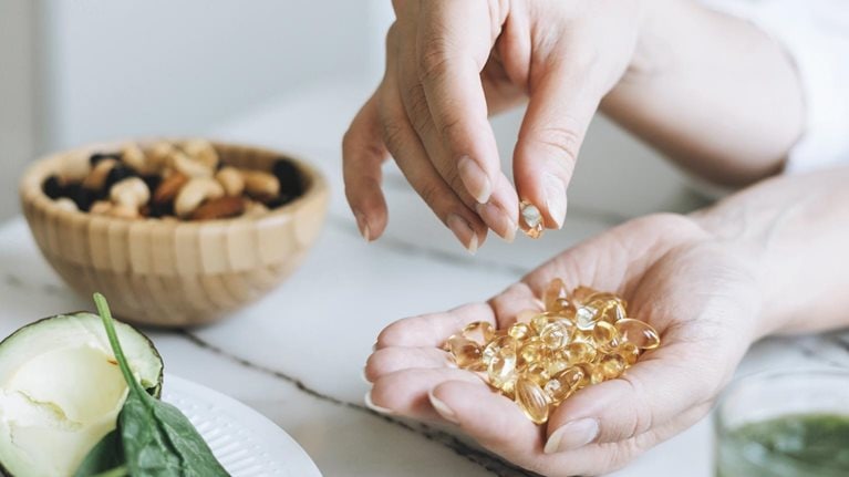
Still feeling good: The US wellness market continues to boom

How to thrive in the global wellness market

Feeling good: The future of the $1.5 trillion wellness market
Suggestions or feedback?
MIT News | Massachusetts Institute of Technology
- Machine learning
- Social justice
- Black holes
- Classes and programs
Departments
- Aeronautics and Astronautics
- Brain and Cognitive Sciences
- Architecture
- Political Science
- Mechanical Engineering
Centers, Labs, & Programs
- Abdul Latif Jameel Poverty Action Lab (J-PAL)
- Picower Institute for Learning and Memory
- Lincoln Laboratory
- School of Architecture + Planning
- School of Engineering
- School of Humanities, Arts, and Social Sciences
- Sloan School of Management
- School of Science
- MIT Schwarzman College of Computing
A prosthesis driven by the nervous system helps people with amputation walk naturally
Press contact :.
Previous image Next image
State-of-the-art prosthetic limbs can help people with amputations achieve a natural walking gait, but they don’t give the user full neural control over the limb. Instead, they rely on robotic sensors and controllers that move the limb using predefined gait algorithms.
Using a new type of surgical intervention and neuroprosthetic interface, MIT researchers, in collaboration with colleagues from Brigham and Women’s Hospital, have shown that a natural walking gait is achievable using a prosthetic leg fully driven by the body’s own nervous system. The surgical amputation procedure reconnects muscles in the residual limb, which allows patients to receive “proprioceptive” feedback about where their prosthetic limb is in space.
In a study of seven patients who had this surgery, the MIT team found that they were able to walk faster, avoid obstacles, and climb stairs much more naturally than people with a traditional amputation.

“This is the first prosthetic study in history that shows a leg prosthesis under full neural modulation, where a biomimetic gait emerges. No one has been able to show this level of brain control that produces a natural gait, where the human’s nervous system is controlling the movement, not a robotic control algorithm,” says Hugh Herr, a professor of media arts and sciences, co-director of the K. Lisa Yang Center for Bionics at MIT, an associate member of MIT’s McGovern Institute for Brain Research, and the senior author of the new study.
Patients also experienced less pain and less muscle atrophy following this surgery, which is known as the agonist-antagonist myoneural interface (AMI). So far, about 60 patients around the world have received this type of surgery, which can also be done for people with arm amputations.
Hyungeun Song, a postdoc in MIT’s Media Lab, is the lead author of the paper , which appears today in Nature Medicine .
Sensory feedback
Most limb movement is controlled by pairs of muscles that take turns stretching and contracting. During a traditional below-the-knee amputation, the interactions of these paired muscles are disrupted. This makes it very difficult for the nervous system to sense the position of a muscle and how fast it’s contracting — sensory information that is critical for the brain to decide how to move the limb.
People with this kind of amputation may have trouble controlling their prosthetic limb because they can’t accurately sense where the limb is in space. Instead, they rely on robotic controllers built into the prosthetic limb. These limbs also include sensors that can detect and adjust to slopes and obstacles.
To try to help people achieve a natural gait under full nervous system control, Herr and his colleagues began developing the AMI surgery several years ago. Instead of severing natural agonist-antagonist muscle interactions, they connect the two ends of the muscles so that they still dynamically communicate with each other within the residual limb. This surgery can be done during a primary amputation, or the muscles can be reconnected after the initial amputation as part of a revision procedure.
“With the AMI amputation procedure, to the greatest extent possible, we attempt to connect native agonists to native antagonists in a physiological way so that after amputation, a person can move their full phantom limb with physiologic levels of proprioception and range of movement,” Herr says.
In a 2021 study , Herr’s lab found that patients who had this surgery were able to more precisely control the muscles of their amputated limb, and that those muscles produced electrical signals similar to those from their intact limb.
After those encouraging results, the researchers set out to explore whether those electrical signals could generate commands for a prosthetic limb and at the same time give the user feedback about the limb’s position in space. The person wearing the prosthetic limb could then use that proprioceptive feedback to volitionally adjust their gait as needed.
In the new Nature Medicine study, the MIT team found this sensory feedback did indeed translate into a smooth, near-natural ability to walk and navigate obstacles.
“Because of the AMI neuroprosthetic interface, we were able to boost that neural signaling, preserving as much as we could. This was able to restore a person's neural capability to continuously and directly control the full gait, across different walking speeds, stairs, slopes, even going over obstacles,” Song says.
A natural gait
For this study, the researchers compared seven people who had the AMI surgery with seven who had traditional below-the-knee amputations. All of the subjects used the same type of bionic limb: a prosthesis with a powered ankle as well as electrodes that can sense electromyography (EMG) signals from the tibialis anterior the gastrocnemius muscles. These signals are fed into a robotic controller that helps the prosthesis calculate how much to bend the ankle, how much torque to apply, or how much power to deliver.
The researchers tested the subjects in several different situations: level-ground walking across a 10-meter pathway, walking up a slope, walking down a ramp, walking up and down stairs, and walking on a level surface while avoiding obstacles.
In all of these tasks, the people with the AMI neuroprosthetic interface were able to walk faster — at about the same rate as people without amputations — and navigate around obstacles more easily. They also showed more natural movements, such as pointing the toes of the prosthesis upward while going up stairs or stepping over an obstacle, and they were better able to coordinate the movements of their prosthetic limb and their intact limb. They were also able to push off the ground with the same amount of force as someone without an amputation.
“With the AMI cohort, we saw natural biomimetic behaviors emerge,” Herr says. “The cohort that didn’t have the AMI, they were able to walk, but the prosthetic movements weren’t natural, and their movements were generally slower.”
These natural behaviors emerged even though the amount of sensory feedback provided by the AMI was less than 20 percent of what would normally be received in people without an amputation.
“One of the main findings here is that a small increase in neural feedback from your amputated limb can restore significant bionic neural controllability, to a point where you allow people to directly neurally control the speed of walking, adapt to different terrain, and avoid obstacles,” Song says.
“This work represents yet another step in us demonstrating what is possible in terms of restoring function in patients who suffer from severe limb injury. It is through collaborative efforts such as this that we are able to make transformational progress in patient care,” says Matthew Carty, a surgeon at Brigham and Women’s Hospital and associate professor at Harvard Medical School, who is also an author of the paper.
Enabling neural control by the person using the limb is a step toward Herr’s lab’s goal of “rebuilding human bodies,” rather than having people rely on ever more sophisticated robotic controllers and sensors — tools that are powerful but do not feel like part of the user’s body.
“The problem with that long-term approach is that the user would never feel embodied with their prosthesis. They would never view the prosthesis as part of their body, part of self,” Herr says. “The approach we’re taking is trying to comprehensively connect the brain of the human to the electromechanics.”
The research was funded by the MIT K. Lisa Yang Center for Bionics and the Eunice Kennedy Shriver National Institute of Child Health and Human Development.

Previous item Next item
Share this news article on:
Press mentions, the guardian.
MIT scientists have conducted a trial of a brain controlled bionic limb that improves gait, stability and speed over a traditional prosthetic, reports Hannah Devlin for The Guardian . Prof. Hugh Herr says with natural leg connections preserved, patients are more likely to feel the prosthetic as a natural part of their body. “When the person can directly control and feel the movement of the prosthesis it becomes truly part of the person’s anatomy,” Herr explains.
The Economist
Using a new surgical technique, MIT researchers have developed a bionic leg that can be controlled by the body’s own nervous system, reports The Economist . The surgical technique “involved stitching together the ends of two sets of leg muscles in the remaining part of the participants’ legs,” explains The Economist . “Each of these new connections forms a so-called agonist-antagonist myoneural interface, or AMI. This in effect replicates the mechanisms necessary for movement as well as the perception of the limb’s position in space. Traditional amputations, in contrast, create no such pairings.”
The Boston Globe
Researchers at MIT and Brigham and Women’s Hospital have created a new surgical technique and neuroprosthetic interface for amputees that allows a natural walking gait driven by the body’s own nervous system, reports Adam Piore for The Boston Globe . “We found a marked improvement in each patient’s ability to walk at normal levels of speed, to maneuver obstacles, as well as to walk up and down steps and slopes," explains Prof. Hugh Herr. “I feel like I have my leg — like my leg hasn’t been amputated,” shares Amy Pietrafitta, a participant in the clinical trial testing the new approach.
Researchers at MIT have developed a novel surgical technique that could “dramatically improve walking for people with below-the-knee amputations and help them better control their prosthetics,” reports Timmy Broderick for STAT . “With our patients, even though their limb is made of titanium and silicone, all these various electromechanical components, the limb feels natural, and it moves naturally, without even conscious thought," explains Prof. Hugh Herr.
Financial Times
A new surgical approach developed by MIT researchers enables a bionic leg driven by the body’s nervous system to restore a natural walking gait more effectively than other prosthetic limbs, reports Clive Cookson for the Financial Times . “The approach we’re taking is trying to comprehensively connect the brain of the human to the electro-mechanics,” explains Prof. Hugh Herr.
The Washington Post
A new surgical procedure and neuroprosthetic interface developed by MIT researchers allows people with amputations to control their prosthetic limbs with their brains, “a significant scientific advance that allows for a smoother gait and enhanced ability to navigate obstacles,” reports Lizette Ortega for The Washington Post . “We’re starting to get a glimpse of this glorious future wherein a person can lose a major part of their body, and there’s technology available to reconstruct that aspect of their body to full functionality,” explains Prof. Hugh Herr.
Related Links
- Biomechatronics Group
- McGovern Institute
- K. Lisa Yang Center for Bionics
Related Topics
- Assistive technology
- Prosthetics
- Neuroscience
- School of Architecture and Planning
Related Articles
Magnetic sensors track muscle length

New surgery may enable better control of prosthetic limbs
Making prosthetic limbs feel more natural
More mit news.

Q&A: What past environmental success can teach us about solving the climate crisis
Read full story →

Marking a milestone: Dedication ceremony celebrates the new MIT Schwarzman College of Computing building

Machine learning and the microscope

Reasoning skills of large language models are often overestimated

MIT SHASS announces appointment of new heads for 2024-25

When to trust an AI model
- More news on MIT News homepage →
Massachusetts Institute of Technology 77 Massachusetts Avenue, Cambridge, MA, USA
- Map (opens in new window)
- Events (opens in new window)
- People (opens in new window)
- Careers (opens in new window)
- Accessibility
- Social Media Hub
- MIT on Facebook
- MIT on YouTube
- MIT on Instagram
Paper Leak Serious Issue, Urgent Need for Structural Reforms in Exam System: Delhi University VC
Last Updated: July 14, 2024, 10:10 IST
New Delhi, India

Delhi University Vice Chancellor Yogesh Singh. (Pic credit: Screengrab from ANI video.)
Delhi University VC Yogesh Singh said the Centre must act against those involved in "rigging” of exams and expressed hope that the CBI inquiry into the alleged paper leak of NEET-UG and UGC-NET exams will yield a positive result
Delhi University Vice-Chancellor Yogesh Singh on Friday said paper leak in various competitive exams is a serious issue and there is an urgent need for structural reforms in the examination system to prevent such irregularities.
He said the Centre must act against those involved in “rigging” of exams and expressed hope that the CBI inquiry into the alleged paper leak of NEET-UG and UGC-NET exams will yield a positive result.
In an interview with PTI, Singh exuded confidence that things will improve soon as the government is tackling the issue with seriousness.
“There are criminal gangs in the country that try to leak exam papers. First, action must be taken against those gangs. Second, structural reforms within the examination system are required which (should) evolve with time,” he said.
The Centre faces the challenge of eliminating such elements, the vice-chancellor said.
“The CBI inquiry ordered into the incidents of paper leak will yield a positive result and instil a sense of fear in the minds of criminals.
“This is a very serious matter. We need to look at it from a wider perspective… (instead of) just focus(ing) on irregularities in one or two exams. I believe that the situation will improve when reforms are taken into consideration,” Singh said.
Given the seriousness with which the government is tackling the problem, the situation will improve soon, he asserted.
While NEET-UG is under the scanner for several irregularities, including an alleged paper leak, UGC-NET was cancelled after the Ministry of Education received inputs that the integrity of the exam was compromised. Two other exams — CSIR-UGC NET and NEET PG — were cancelled as a pre-emptive step.
The National Testing Agency (NTA), which conducts these exams, later announced fresh dates for some of these exams.
The Centre has set up a seven-member panel headed by former ISRO chief R Radhakrishnan to make recommendations on reforms in the examination process, enhancing data security protocols, and reviewing the structure and operations of the NTA.
The NTA also conducts the Common University Entrance Test (CUET) for admission to central universities.
The results for CUET-UG exam are yet to be released by the NTA. As a result, admissions in many central universities, including DU, have been delayed and their academic calendars will be cramped.
Stay ahead with all the exam results updates on News18 Website .
- delhi university
- DU admissions

IMAGES
VIDEO
COMMENTS
Lotions can chill, calm, or protect locally [1]. A herbal body lotion is a. liquid c omposition applied to the skin to improve aesthetics. Lotions remove sebum and. cleanse the skin. This chemical ...
Petrolatum is the most effective classic occlusive moisturizer; a minimum concentration of 5%, can reduce trans-epidermal water loss by more than 98%, with 170-times water vapor loss resistance as compared to olive oil. 16. Lanolin, mineral oil and silicones (eg, dimethicone) can reduce trans-epidermal water loss by 20% to 30%.
Body and hand feet moisturizers: They are mostly aimed at prevention as well as treatment of dry skin, eczema, and xerosis. They are dispensed in the form of lotions, creams, and mousse. Some specialized products aims include cellulite firming, bronzing, and minimizing the signs of aging.[ 5 ]
containing herbal lotion protects your skin from harmful pollution effects, smoothens the skin and gives a freshness feel to the skin.The skin is the body 's. largest organ, made of water, protein ...
Abstract. Moisturizers provide functional skin benefits, such as making the skin smooth and soft, increasing skin hydration, and improving skin optical characteristics; however, moisturizers also function as vehicles to deliver ingredients to the skin. These ingredients may be vitamins, botanical antioxidants, peptides, skin-lightening agents ...
Stability studies of the lotion showed that the lotion was stable after three months. Journal of Institute of Science and Technology Volume 21, Issue 1, August 2016, Page: 148-156</p View
All lotion formulations were prepared by mixing the ingredients as given in the Table 1. Essentially, 2 g of DDA was dissolved in 20 mL of ethanol and this solution was added to the 20 mL of phosphate buffered saline containing 980 mg of carbomer. These were mixed for 30 minutes until a clear solution was obtained.
nterest in utilizing VCO for cosmetic formulations. A study was conducted at the Regional Agricultural Research Station, Pilicode for developing a body lotion using virgin coconut oil as the base, along with Aloe ve. a, bee wax, cocoa butter, and fragrance principles. Considering the results obtained in sensory analysis, a combination of body ...
containing herbal lotion protects your skin from harmful pollution effects, smoothens the skin and gives a freshness feel to the skin.The skin is the body's largest organ, made of water, protein, fats and minerals[5]. Your skin protects your body from germs and regulates body temperature. Nerves in the skin help you feel
The body lotion is made in the form of an oil in water (m/a) and made in 4 formulas the concentration of the extract 0.5 %, 1%, and 2%. The physical test of body lotion extract of Labisia pumila is organoleptic, homogeneity, pH, viscosity, the spread, and the attaching, the results of testing in analysis using SPSS with Kruskal-Wallis and Mann ...
There is a growing body of literature that recognizes the importance of moisturizers. It is essential for a wide range of fields, such as cosmetics and pharmacy [1]. Moisturizers are very popular dermatological products prescribed due to their proven efficiency to prevent and treat various dermatological conditions [2,3].
This paper presents the preliminary report of the 2018 season of the expedition of the University of Pisa in the area of tomb M.I.D.A.N.05, at Dra Abu el-Naga (Theban Necropolis). ... Evaluation of herbal body lotion Evaluation research is defined as a form of disciplined and systematic inquiry that is carried out to arrive at an assessment or ...
The main findings obtained from this study are summarized as follows: (1) Body cream and body lotion exhibit a finite magnitude of yield stress. This feature is directly related to the primary (initial) skin feel that consumers usually experience during actual usage. (2) Body cream and body lotion exhibit a pronounced shear-thinning behavior.
Background Use of skin personal care products on a regular basis is nearly ubiquitous, but their effects on molecular and microbial diversity of the skin are unknown. We evaluated the impact of four beauty products (a facial lotion, a moisturizer, a foot powder, and a deodorant) on 11 volunteers over 9 weeks. Results Mass spectrometry and 16S rRNA inventories of the skin revealed decreases in ...
Virgina Coconut Oil is containing saturated fatty acid compound such as lauric and oleic which can soften dry and rough skin. While the natural ingridients of black tea extract, secang wood and telang flower contain polyphenol compounds which have antioxidant activity, they also have attractive colors making them suitable as natural dyes for cosmetic ingredients.
This research work unimpeachably exposed the several challenges associated with allopathic lotion such as side effect, high cost and sensitivity. The herbal lotion of crude drugs with the unique properties and prepared by simple W/O methods and less equipment are required. In which the research of study was concluded that
A portion of lotion was applied on the forearms of 6 volunteers and left for 20 minutes. After 20 minutes any kind of irritation if occurred was noted. 13. Washability Test A portion of lotion was applied over the skin of hand and allowed to flow under the force of flowing tap water for 10 minutes. The time when the lotion completely removed ...
Herein, a number of typical yield stress fluids including carbopol gel, 56-58 laponite suspension, 59 polysaccharide solutions of xanthan gum 3 and welan gum, 60 gel particle suspension, 61 ...
The data published suggests that the tested body lotion provides the skin with nutrients to boost the production of the range of essential skin lipids needed for well-nourished, well-moisturised and healthier skin. Mapping the microbiome. The research team was also interested in how the product changed the skin microbiome after application.
DOI: 10.33545/26647222.2023.v5.i2a.41 Corpus ID: 261928197; Pharmaceutical assessment of body lotion: A herbal formulation and its potential benefits @article{Mishra2023PharmaceuticalAO, title={Pharmaceutical assessment of body lotion: A herbal formulation and its potential benefits}, author={Sunil Mishra and Dr. Shashank Tiwari and Kartikay Prakash and Prachi Jaiswal and Harsh Rajpoot ...
ar, pigmentation, redness and itching of the skin.This body lotion was formulated and evaluated by different evaluation parameters such as pH, viscosity, spreadabilty, physical appearance and Irritancytest Stability testing for prepared formulation was performed by storing it at different tempe. ture condition for time period of 24h for 1 week ...
Building upon last year's research, several pockets of growth in the wellness space are emerging. Increasing consumer interest, technological breakthroughs, product innovation, and an increase in chronic illnesses have catalyzed growth in these areas. Women's health . Historically, women's health has been underserved and underfunded ...
Capital markets regulator Sebi on Friday said it has recognised BSE Ltd as a supervisory body for research analysts and investment advisers to oversee their management and administration. In its circular, Sebi said, "BSE Ltd has been granted recognition under regulation 14 of the RA regulations ...
The main findings obtained from this study are summarized as follows: (1) Body cream and body lotion exhibit a finite magnitude of yield stress. This feature is directly related to the primary ...
"This is the first prosthetic study in history that shows a leg prosthesis under full neural modulation, where a biomimetic gait emerges. No one has been able to show this level of brain control that produces a natural gait, where the human's nervous system is controlling the movement, not a robotic control algorithm," says Hugh Herr, a professor of media arts and sciences, co-director ...
Delhi University Vice-Chancellor Yogesh Singh on Friday said paper leak in various competitive exams is a serious issue and there is an urgent need for structural reforms in the examination system to prevent such irregularities. ... Research Analysts: BSE Now Your Supervisory Body, Gets Sebi Recognition ...
Untuk evaluasi karakteristik fisik lotion menunjukkan bahwa lotion yang diperoleh t, memiliki warna hijau, homogen; pH lotion 8,02-6,98; viskositas lotion 3545-4053 cps, daya lekat rata-rata 3,23 ...