- Previous Article
- Next Article

Utilization of corn husk for tissue papermaking
- Article contents
- Figures & tables
- Supplementary Data
- Peer Review
- Reprints and Permissions
- Cite Icon Cite
- Search Site
Natalia Suseno , Marisca E. Gondokesumo , Puspita R. Permatasari; Utilization of corn husk for tissue papermaking. AIP Conf. Proc. 11 November 2021; 2338 (1): 040019. https://doi.org/10.1063/5.0067417
Download citation file:
- Ris (Zotero)
- Reference Manager
The demand of tissue papers is increasing with the population increase. This will definitely increase the need of wood fibers as the main raw material. However, due to the wood shortages, there have been many attempts to use non- wood fibers as substitutes for papermaking. In Indonesia, corn production has gradually increased for the last 5 years, hence it also has an impact on the raising in the amount of corn husk waste. Corn husk has a high cellulose content which suitable to be used as a raw material for tissue papermaking. In this experiment, soda pulping process was conducted to remove out lignin. The resulting tissue paper will be added with additives that have antimicrobial properties of chitosan and mangosteen peel for the purpose of increasing the tensile strength or absorption of water. The aim of this research is to study the effect of depending variables (temperature and NaOH concentration) on chemical composition (cellulose and lignin content), and physical properties including water absorption and tensile strength.The research was started with the initial process of removing the lignin content in the pulp by pretreating delignification using the sodium hydroxide (NaOH) process with several variations in concentration (4–10%), and temperature (60–90°C) for 1.5 hours. To obtain tissue with a good physical condition, it has been influenced by the optimum chemical composition containing high cellulose and low lignin content, high tensile strength and water absorption. The optimum conditions for tissue paper in this study were at 90°C and 4% of NaOH concentration. The next step will be to vary the composition of the additive in order to obtain the effect of physical properties (tensile strength and water absorption).
Sign in via your Institution
Citing articles via, publish with us - request a quote.

Sign up for alerts
- Online ISSN 1551-7616
- Print ISSN 0094-243X
- For Researchers
- For Librarians
- For Advertisers
- Our Publishing Partners
- Physics Today
- Conference Proceedings
- Special Topics
pubs.aip.org
- Privacy Policy
- Terms of Use
Connect with AIP Publishing
This feature is available to subscribers only.
Sign In or Create an Account
New Research
This “Tissue” Paper Is Made From Real Tissue
Made from powdered organs, the flexible paper could be used as a sophisticated bandage during surgery
Jason Daley
Correspondent
/https://tf-cmsv2-smithsonianmag-media.s3.amazonaws.com/filer/8e/ec/8eec902a-29d2-436c-98cf-1487cfb51937/origami_.jpg "tissue paper research")
When Adam Jakus was a postdoc at Northwestern University he accidentally spilled some “ink” he'd created from powdered ovaries intended for 3-D printing. Before he could wipe up the mess, it solidified into a thin, paper-like sheet, reports Charles Q. Choi at LiveScience . That led to a lab-bench epiphany.
“When I tried to pick it up, it felt strong,” Jakus says in a press release . “I knew right then I could make large amounts of bioactive materials from other organs. The light bulb went on in my head.”
Jakus, along with the same team that developed a 3-D printed mouse ovary earlier this year, began experimenting with the concept. According to a video , they began collecting pig and cow organs from a local butcher shop, including livers, kidneys, ovaries, uteruses, hearts and muscle tissue.
The team then used a solution to strip the cells from the tissues, leaving behind a the scaffolding material of collagen proteins and carbohydrates. After freeze-drying the matrix, they powdered it and mixed it with materials that allowed them to form it into thin sheets. The research appears in the journal Advanced Functional Materials .
“We’ve created a material we call 'tissue papers' that’s very thin, like phyllo dough, made up of biological tissues and organs,” says Ramille Shah, head of the lab where the research took place, in the video. “We can switch out the tissue we use to make the tissue paper—whether that be derived from liver or muscle or even ovary. We can switch it out very easily and make a paper out of any tissue or organ.”
According to the press release, the material is very paper-like and can be stacked in sheets. Jakus even folded some into origami cranes. But the tissue paper’s most important property is that it is biocompatible and allows for cellular growth. For instance, the team seeded the paper with stem cells, which attached to the matrix and grew over four weeks.
That means the material could potentially be useful in surgery, since paper made of muscle tissue could be used as a sophisticated Band-Aid to repair injured organs. “They're easy to store, fold, roll, suture and cut, like paper," Jakus tells Choi. “Their flat, flexible nature is important if doctors want to shape and manipulate them in surgical situations.”
Northwestern reproductive scientist Teresa Woodruff was also able to grow ovary tissue from cows on the paper, which eventually began producing hormones. In the press release, she explains that a strip of the hormone-producing tissue paper could be implanted, possibly under the arm, of girls who have lost their ovaries due to cancer treatments to help them reach puberty.
The idea of using extracellular matrices, hydrogels or other material as a scaffolding to bioprint organs like hearts and kidneys is being investigated by labs around the world. In 2015, a Russian team claimed they printed a functional mouse thyroid . And this past April, researchers were able to b ioprint a patch derived from human heart tissue that they used to repair the heart of a mouse.
Get the latest stories in your inbox every weekday.
Jason Daley | | READ MORE
Jason Daley is a Madison, Wisconsin-based writer specializing in natural history, science, travel, and the environment. His work has appeared in Discover , Popular Science , Outside , Men’s Journal , and other magazines.

An official website of the United States government
The .gov means it’s official. Federal government websites often end in .gov or .mil. Before sharing sensitive information, make sure you’re on a federal government site.
The site is secure. The https:// ensures that you are connecting to the official website and that any information you provide is encrypted and transmitted securely.
- Publications
- Account settings
- My Bibliography
- Collections
- Citation manager
Save citation to file
Email citation, add to collections.
- Create a new collection
- Add to an existing collection
Add to My Bibliography
Your saved search, create a file for external citation management software, your rss feed.
- Search in PubMed
- Search in NLM Catalog
- Add to Search
Experimental dataset supporting the physical and mechanical characterization of industrial base tissue papers
Affiliations.
- 1 Fibre Materials and Environmental Technologies Research Unit (FibEnTech-UBI), Universidade da Beira Interior, Rua Marquês d'Ávila e Bolama, Covilhã 6201-001, Portugal.
- 2 Forest and Paper Research Institute (RAIZ), R. José Estevão, Eixo, Aveiro 3800-783, Portugal.
- PMID: 33145380
- PMCID: PMC7593524
- DOI: 10.1016/j.dib.2020.106434
Tissue paper is defined by its physical and mechanical properties, namely: high softness, low grammage, high bulk and high liquid absorption capacity. It is expected that the production of tissue paper will continue to grow, which increases the importance of better understanding the processes involved in its production as well as its optimization [1]. The experimental data presented in this article, are the physical-mechanical characterization of a group of 13 industrial base tissue papers, which were collected at the end of the tissue paper machine on Portuguese factories. These samples vary in grammage, composition and creping [2], enabling a later evaluation of the crepe type [3] and its relationship with the final properties of the tissue paper.
Keywords: Absorption capacity; Fiber morphology; Industrial base tissue paper; Mechanical characterization; Structural properties; Tissue softness.
© 2020 The Author(s).
PubMed Disclaimer
Conflict of interest statement
The authors declare that they have no known competing financial interests or personal relationships which have, or could be perceived to have, influenced the work reported in this article.
Similar articles
- FEM Analysis Validation of Rubber Hardness Impact on Mechanical and Softness Properties of Embossed Industrial Base Tissue Papers. Vieira JC, Mendes AO, Ribeiro ML, Vieira AC, Carta AM, Fiadeiro PT, Costa AP. Vieira JC, et al. Polymers (Basel). 2022 Jun 18;14(12):2485. doi: 10.3390/polym14122485. Polymers (Basel). 2022. PMID: 35746060 Free PMC article.
- Challenges in computational materials modelling and simulation: A case-study to predict tissue paper properties. Morais FP, Curto JMR. Morais FP, et al. Heliyon. 2022 May 2;8(5):e09356. doi: 10.1016/j.heliyon.2022.e09356. eCollection 2022 May. Heliyon. 2022. PMID: 35540931 Free PMC article.
- Embossing Pressure Effect on Mechanical and Softness Properties of Industrial Base Tissue Papers with Finite Element Method Validation. Vieira JC, Mendes AO, Ribeiro ML, Vieira AC, Carta AM, Fiadeiro PT, Costa AP. Vieira JC, et al. Materials (Basel). 2022 Jun 18;15(12):4324. doi: 10.3390/ma15124324. Materials (Basel). 2022. PMID: 35744382 Free PMC article.
- Mechanical Metamaterials on the Way from Laboratory Scale to Industrial Applications: Challenges for Characterization and Scalability. Fischer SCL, Hillen L, Eberl C. Fischer SCL, et al. Materials (Basel). 2020 Aug 14;13(16):3605. doi: 10.3390/ma13163605. Materials (Basel). 2020. PMID: 32824029 Free PMC article. Review.
- Organic and mechanical properties of Cervidae antlers: a review. Picavet PP, Balligand M. Picavet PP, et al. Vet Res Commun. 2016 Dec;40(3-4):141-147. doi: 10.1007/s11259-016-9663-8. Epub 2016 Sep 12. Vet Res Commun. 2016. PMID: 27618827 Review.
- Enhanced water absorption of tissue paper by cross-linking cellulose with poly(vinyl alcohol). Ferreira ACS, Aguado R, Bértolo R, Carta AMMS, Murtinho D, Valente AJM. Ferreira ACS, et al. Chem Zvesti. 2022;76(7):4497-4507. doi: 10.1007/s11696-022-02188-y. Epub 2022 Apr 8. Chem Zvesti. 2022. PMID: 35431412 Free PMC article.
- Raunio Simulation of creping pattern in tissue paper. Nordic Pulp Paper Res. J. 2012;27:375–381. doi: 10.3183/NPPRJ-2012-27-02-p375-381. - DOI
- Anukul P., Khantayanuwong S., Somboon P. Development of laboratory wet creping method to evaluate and control pulp quality for tissue. TAPPI J. 2015;14:7.
- Ramasubramanian M.K., Shmagin D.L. An experimental investigation of the creping process in low-density paper manufacturing. J. Manuf. Sci. Eng. 2000;122:576. doi: 10.1115/1.1285908. - DOI
- J.C. Vieira, A. de O. Mendes, A.M. Carta, E. Galli, P.T. Fiadeiro, A.P. Costa, Impact of Embossing on Liquid Absorption of Toilet Tissue Papers. 15 (2020) 3888–3898. doi:10.15376/biores.15.2.3888-3898. - DOI
- ISO 12625-6:2005 Tissue Paper and Tissue Products - Part 6: Determination of Grammage (2005).
LinkOut - more resources
Full text sources.
- Elsevier Science
- Europe PubMed Central
- PubMed Central

- Citation Manager
NCBI Literature Resources
MeSH PMC Bookshelf Disclaimer
The PubMed wordmark and PubMed logo are registered trademarks of the U.S. Department of Health and Human Services (HHS). Unauthorized use of these marks is strictly prohibited.
Please fill the all required fields....!!
World of TISSUES

The tissues sector has boomed over the last few years. With a move to more luxurious tissue paper and ultra absorbent paper towels the industry has been able to increase the tissue prices and create new brands to retain consumers. Add to this growing market in developing countries and a drive to innovate, and there are very positive signals for the future of tissues. The tissue segment has become one of the most important segments of the paper industry in developed countries. It produces one of the most highly valued and appreciated, though not necessarily talked about, paper products.
Tissue paper or simply tissue is a lightweight paper or, light crêpe paper. Tissue can be made both from virgin and recycled paper pulp. Tissue papers are the most common thing that we use in our daily life from cleaning, dusting, wrapping and personal use.
As far as the history of tissue paper industry goes, although paper had been known as a wrapping and padding material in China since the 2nd century BC, the first use of toilet paper in human history dates back to the 6th century AD, in early medieval China. If we could travel back in time to 1391, we would encounter a Chinese emperor who demanded the first paper sheets sliced to be placed in his outhouse. The first “official” toilet paper was introduced in China measuring a whopping 2 ft X 3 ft each.
An important move towards the production and distribution of modern toilet tissue paper came from a teacher in Philadelphia in 1907. Concerned about a mild cold epidemic in her classroom, she blamed it on the fact that all students used the same cloth towel. She proceeded to cut up paper into squares to be used by her class as individual towels, a revolutionary idea. Arthur Scott of Scott Paper Company heard about this teacher and decided he would try to sell the carload of paper. He perforated the thick paper into small towel-size sheets and sold them as disposable paper towels. Later he renamed the product Sani-Towel and sold them to hotels, restaurants, and railroad stations for use in public washrooms.
In 1931, Scott introduced the first paper towel for the kitchen and created a whole new grocery category. He made perforated rolls of "towels" thirteen inches wide and eighteen inches long. That is how paper towels were born. It was to take many years, however, before they gained acceptance and replaced cloth towels for kitchen use.
Production of the tissue paper
Tissue paper is produced on a paper machine that has a single large steam heated drying cylinder (yankee dryer) fitted with a hot air hood. The raw material is paper pulp. The yankee cylinder is sprayed with adhesives to make the paper stick. Creping is done by the yankee's “doctor blade” that is scraping the dry paper off the cylinder surface. The crinkle (creping) is controlled by the strength of the adhesive, geometry of the doctor blade, speed difference between the yankee and final section of the paper machine and paper pulp characteristics.
The highest water absorbing applications are produced with a through air drying (TAD) process. These papers contain high amounts of NBSK and CTMP. This gives a bulky paper with high wet tensile strength and good water holding capacity. The TAD process uses about twice the energy compared with conventional drying of paper.
The properties are controlled by pulp quality, creping and additives (both in base paper and as coating). The wet strength is often an important parameter for tissue paper.
Applications of tissue paper
Tissue paper has number of applications and they come in number of varieties such as hygienic tissues, facial tissues, paper towels, wrapping tissues, toilet tissues, table napkins etc.
- Facial tissue has been used for centuries in Japan, in the form of washi (??) or Japanese tissue, as described in this 17th-century European account of the voyage of Hasekura Tsunenaga. In 1924 facial tissue as it is known today was first introduced by Kimberly-Clark as Kleenex. It was invented as a means to remove cold cream. Kimberly-Clark also introduced pop-up, colored, printed, pocket, and 3-ply facial tissues.
- Paper towels are the second largest application for tissue paper in the consumer sector. This type of paper has usually a basis weight of 20 to 24 g/m2. Normally such paper towels are two-ply. This kind of tissue can be made from 100% chemical pulp to 100% recycled fibre or a combination of the two. Normally, some long fibre chemical pulp is included to improve strength. In 1951, William E. Corbin, Henry Chase (scientist), and Harold Titus began experimenting with paper towels in the Research and Development building of the Brown Company in Berlin, New Hampshire. By 1922, Corbin perfected their product and began mass-producing it at the Cascade Mill on the Berlin/Gorham line. This product was called Nibroc Paper Towels (Corbin spelled backwards). In 1931, the Scott Paper Company of Philadelphia, Pennsylvania introduced their paper towel for kitchens. They are now the leader of the manufacture of paper towels.
- W rapping tissue is a type of thin, translucent paper used for wrapping presents and cushioning fragile items.
- Table napkin, or face towel (also in Canada, the United Kingdom, Australia, New Zealand and South Africa: serviette) is a rectangle of cloth used at the table for wiping the mouth and fingers while eating. In the United Kingdom and Canada both terms, serviette and napkin, are used. In certain places, serviettes are those made of paper whereas napkins are made of cloth.
- Rolls of toilet paper have been available since the end of the 19th century. Today, more than 20 billion rolls of toilet tissue are used each year in Western Europe. Joseph Gayetty is widely credited with being the inventor of modern commercially available toilet paper in the United States.
Gayetty's paper, first introduced in 1857, was available as late as the 1920s. Gayetty's Medicated Paper was sold in packages of flat sheets, watermarked with the inventor's name. Seth Wheeler of Albany, New York, obtained the earliest United States patents for toilet paper and dispensers, the types of which eventually were in common usage in that country, in 1883. Moist toilet paper was first introduced in the United Kingdom by Andrex in the 1990s and in the United States by Kimberly-Clark in 2001 (in lieu of bidets which are rare in those countries).
With so many varieties available, tissue industry is growing leaps and bounds. Out of the world's estimated production of 21 million tonnes of tissue, Europe produces approximately 6 million tonnes tissue every year .
The European tissue market is worth approximately 10 billion Euros annually and is growing at a rate of around 3%. The European market represents around 23% of the global market. In North America, people are consuming around three times as much tissue as in Europe. In Europe, the industry is represented by The European Tissue Symposium (ETS), a trade association. The members of ETS represent the majority of tissue paper producers throughout Europe and about 90% of total European tissue production. ETS was founded in 1971 and is based in Brussels since 1992.
Finally, the paper tissue industry, along with the rest of the paper manufacturing sector, has worked hard to minimise its impact on the environment. Recovered fibres now represent some 46.5% of the paper industry’s raw materials. The industry relies heavily on biofuels (about 50% of its primary energy) and it is highly energy-efficient. EDANA, the trade body for the non-woven absorbent hygiene products industry (which includes products such as household wipes for use in the home) has reported annually on the industry’s environmental performance since 2005. The industry’s impact on the environment is in fact, relatively small.
All in all, the tissue sector continues to be a growth industry worldwide, offering new opportunities for companies to expand. Recently, there has been a great deal of interest, in particular, in the use of recovered fibres to manufacture new tissue paper products. However, whether this is actually better for the environment than using new fibres is open to question.
Quick Links
Related Articles

Pilot Paper Machine

Nanotechnology in the pulp and paper industry

About: Devid - Head of Sales
Cras sit amet nibh libero, in gravida nulla. Nulla vel metus scelerisque ante sollicitudin. Cras purus odio, vestibulum in vulputate at, tempus viverra turpis. Fusce condimentum nunc ac nisi vulputate fringilla. Donec lacinia congue felis in faucibus.
Top Viewed Articles
Publish your article.
Thank you for your interest in publishing article with Packaging-Labelling. Our client success team member will get in touch with you shortly to take this ahead.
you're here, check out our informative and insightful article. Happy Surfing!
Client Success Team (CRM),
- Architecture and Design
- Asian and Pacific Studies
- Business and Economics
- Classical and Ancient Near Eastern Studies
- Computer Sciences
- Cultural Studies
- Engineering
- General Interest
- Geosciences
- Industrial Chemistry
- Islamic and Middle Eastern Studies
- Jewish Studies
- Library and Information Science, Book Studies
- Life Sciences
- Linguistics and Semiotics
- Literary Studies
- Materials Sciences
- Mathematics
- Social Sciences
- Sports and Recreation
- Theology and Religion
- Publish your article
- The role of authors
- Promoting your article
- Abstracting & indexing
- Publishing Ethics
- Why publish with De Gruyter
- How to publish with De Gruyter
- Our book series
- Our subject areas
- Your digital product at De Gruyter
- Contribute to our reference works
- Product information
- Tools & resources
- Product Information
- Promotional Materials
- Orders and Inquiries
- FAQ for Library Suppliers and Book Sellers
- Repository Policy
- Free access policy
- Open Access agreements
- Database portals
- For Authors
- Customer service
- People + Culture
- Journal Management
- How to join us
- Working at De Gruyter
- Mission & Vision
- De Gruyter Foundation
- De Gruyter Ebound
- Our Responsibility
- Partner publishers
Your purchase has been completed. Your documents are now available to view.
Online quality evaluation of tissue paper structure on new generation tissue machines
At present, the tissue paper manufacturing is mostly based on the dry crepe technology. During the last decade, the manufacturers have introduced new tissue machines concepts that increase the softness, bulk, and absorption capacity. Such machines produce a strong regular three-dimensional (3D) structure to the sheet before the Yankee cylinder. At present, the quality of the 3D structure is not evaluated, or it is evaluated only subjectively at the mill. This is mostly because of the difficulties to separate reliably the regular 3D pattern from other variations. This paper introduces a frequency analysis based method which separates the surface profile variances in tissue paper to the creping, to the regular 3D pattern and to the residual variation. The 3D surface profiles and their variances were determined online with the photometric stereo method. We show that the introduced analysis method evaluates the variance portions reliably and the results are consistent with the visual perception of the 3D surfaces. In one particular product, the regular 3D pattern explains 74 % of total surface variance; the creping explains 10 % and residual variations 16 %. Furthermore, the creping and residual variances are quite stable over time whereas the variance of the regular 3D pattern fluctuates significantly.
Conflict of interest: The authors do not have any conflicts of interest to declare.
Archer, S., Furman, G. (2005) Embedded sheet Structures-Impact on tissue properties. Tissue World, Nice, France. Search in Google Scholar
Archer, S., Furman, G., Von Drasek, W. (2010) Image analysis to Quantity crepe structure, Proceedings, PaperCon. Tappi, Atlanta, USA. Search in Google Scholar
Boudreau, J. (2013) New methods for evaluation of tissue creping and the importance of coating, paper and adhesion. PhD Thesis, Karlstad University, Karlstad, Sweden. Search in Google Scholar
Frankot, R., Chellappa, R. (1988) A method for enforcing integrability in shape from shading algorithms. IEEE Trans. Pattern Anal. Mach. Intell. 10(4):435–446. 10.1109/34.3909 Search in Google Scholar
Hansson, P., Fransson, P. (2004) Color and shape measurement with a three color photometric stereo system. Appl. Opt. 43(20):3971–3977. 10.1364/AO.43.003971 Search in Google Scholar PubMed
Hollmark, H., Ampulski, R.S. (2004) Measurement of tissue paper softness: A literature review. Nord. Pulp Pap. Res. J. 19(3):345–353. 10.3183/npprj-2004-19-03-p345-353 Search in Google Scholar
Ihalainen, H., Marjanen, K., Mäntylä, M., Kosonen, M. (2012) Developments in camera based on-line measurement of paper. Control systems, New Orleans, USA. Search in Google Scholar
Ikehata, S., Wipf, D., Matsushita, Y., Aizawa, K. (2014) Photometric stereo using sparse Bayesian regression for general diffuse surfaces. IEEE Trans. Pattern Anal. Mach. Intell. 36(18):16–31. 10.1109/TPAMI.2014.2299798 Search in Google Scholar PubMed
Kamps, R.J., Behnke, J.S, Chen, F.-J., Kressner, B.E., Nielsen, J.G. (1994) Method for making soft tissue. U.S. Patent 5743999 A (Filed Jun. 15, 1994). Search in Google Scholar
Klerelid, I., Thomasson, O. (2008) Advantage (TM) NTT: low energy, high quality. Tissue World, Shanghai, China. Search in Google Scholar
Lauri, M., Ihalainen, H. (2010) Measuring periodic patterns in noisy spatial data, Measurement Systems and Process Improvement MSPI 2010-Enbis IMEKO TC 21 Workshop. Teddington, UK. Search in Google Scholar
Mettänen, M. (2010) Measurement of print quality: joint statistical analysis of paper topography and print defects. PhD Thesis, Tampere University of Technology, Tampere, Finland. Search in Google Scholar
Peli, E. (1990) Contrast in Complex Images. J. Opt. Soc. Am. 7(10):2032–2040. 10.1364/JOSAA.7.002032 Search in Google Scholar PubMed
Raunio, J.-P. (2014) Quality Characterization of Tissue and Newsprint Paper based on Image Measurements; Possibilities of On-line Imaging. PhD Thesis, Tampere University of Technology, Tampere, Finland. Search in Google Scholar
Raunio, J.-P., Ritala, R. (2012) Simulation of creping pattern in tissue paper. Nord. Pulp Pap. Res. J. 27(2):375–381. 10.3183/npprj-2012-27-02-p375-381 Search in Google Scholar
Scherb, T., Walkenhaus, H., Herman, J., Silva, L. (2005) Advanced Dewatering System. U.S. Patent 20070256806 A1 (Filed Jan. 19, 2005). Search in Google Scholar
Smith, M.L., Smith L.N. (2005) Dynamic Photometric Stereo - A New Technique for Moving Surface Analysis. Image Vis. Comput. 23(9):841–852. 10.1016/j.imavis.2005.01.007 Search in Google Scholar
Tagare, H.D., deFigueiredo, R.J.P. (1991) A theory of photometric stereo for a class of diffuse non-lambertian surfaces. IEEE Trans. Pattern Anal. Mach. Intell. 13(1):33–52. 10.1109/34.67643 Search in Google Scholar
Welch, P.D. (1967) The use of fast Fourier transform for the estimation of power spectra: a method based on time averaging over short, modified periodograms. IEEE Trans. Audio Electroacoust. 15(2):70–73. 10.1109/TAU.1967.1161901 Search in Google Scholar
Woodham, R.J. (1980) Photometric method for determining surface orientation from multiple images. Opt. Eng. 19(1):139–144. 10.1117/12.7972479 Search in Google Scholar
Woodham, R.J. (1994) Gradient and curvature from photometric-stereo method including local confidence estimation. J. Opt. Soc. Am. 11(11):3050–3068. 10.1364/JOSAA.11.003050 Search in Google Scholar
© 2018 Walter de Gruyter GmbH, Berlin/Boston
- X / Twitter
Supplementary Materials
Please login or register with De Gruyter to order this product.
Journal and Issue
Articles in the same issue.
Perceived Vs Recorded Quality of Tissue Paper: A Thematic Analysis of Online Customer Reviews
- September 2019
- Thesis for: Master of Science in Industrial Engineering and Management
- Advisor: Magnus Berglund

- Linköping University

Discover the world's research
- 25+ million members
- 160+ million publication pages
- 2.3+ billion citations

- Frank Duckhorn

- J RETAILING

- J MARKETING
- Eugene W. Anderson

- Donald R. Lehmann
- INFORM MANAGE-AMSTER
- Chaojiang Wu
- Klaus Mundt
- Michael J. Groves
- Johan Kullander

- Recruit researchers
- Join for free
- Login Email Tip: Most researchers use their institutional email address as their ResearchGate login Password Forgot password? Keep me logged in Log in or Continue with Google Welcome back! Please log in. Email · Hint Tip: Most researchers use their institutional email address as their ResearchGate login Password Forgot password? Keep me logged in Log in or Continue with Google No account? Sign up
Renewable Bioproducts Institute
Pulp, paper, packaging and tissue.
From tree to tissue, or to package, or to stationery, magazine or newspaper or thousands of other products—pulping liberates fibers from woody plants and rearranges them into a consistently formed end products. The science rests in efficient processes that conserve energy and raw materials while producing the desired product.

In the 1950s, the kraft chemical pulping process became the dominant technology for isolating wood fiber from chips. While this produces strong pulp and efficient recovery of raw materials and energy, the yield is below ideal and has an inherent capital intensity for the investor. RBI researchers are developing improvements in next generation pulping to address these challenges.
We are exploring pretreatment options to make fiber separation more efficient, and improving yields. A number of catalysis options, including biochemical methods, are under investigation. Biorefining processes are converting cellulose and lignin into sugars, fuels, and chemical intermediates and feedstocks.
Advanced processes under investigation include modeling tools for evaporation of water from spent pulping liquor and techniques to eliminate equipment fouling. Advanced membrane technologies are expanding capabilities for cost-effective separation of organics and inorganics from spent pulping liquor. This opens up new possibilities for reducing energy intensity and even more efficient reuse of process streams.
Understanding the pathways of erosion and corrosion, and developing means to eliminate them, can save millions in maintenance and replacement costs, as well as lost production. Analysis and testing capabilities include thermodynamic prediction and modeling of corrosion processes and recommendations for improved metallurgy to address the aggressive environments in these applications.
At RBI, we continue to explore the seemingly limitless possibilities of paper. As the successor to the Institute of Paper Chemistry founded in 1929 by leaders of the pulp and paper industry, our mission is to create a body of knowledge and advance the industry. Our researchers are working to understand and manage new ways in which: • molecular structures in fibers can be coaxed to bond (or not) • the structure of a sheet can be formed • moisture, gases, oil and grease can be absorbed or repelled • inks can be accepted or released • formation and de-watering will improve manufacturing and energy efficiency These mechanisms and more are creating inspiration for new products of the 21st century. Operational excellence is our goal. In taking the industry into this new era of technology and application of science, we continue to develop paper-based substrates and biocomposites for applications in electronics used in smart packaging; high-strength, light-weight panels for aviation and automotive uses; advanced technologies for paper, packaging and films; strength and fracture characteristics of paper for packaging; superamphiphobic paper for water and grease repellency; and auxetic papers that expand when stretched. Innovative technologies have resulted in new applications, products and materials from renewable, sustainable forest biomass.
New Product Innovations
RBI continues to address challenges and opportunities in fiber engineering and paper physics to yield a step-change in paper/packaging product performance. For those members who utilize the fundamental knowledge embedded in our faculty and staff, RBI creates a competitive edge and insight into the future of forest products.
In the area of consumer products, technologies such as light dry crepe and through-air drying, non-woven technologies such as spunbond, meltblown and spunlace are included in the expertise of our faculty. The application of biomaterials will be expanded in consumer products within the next few years across the spectrum of disposables and single-use plastics to create a more sustainable offering within this segment of the industry.
Some of the new product innovations in paper board include smart packaging, coating solutions, printed electronics, renewable binders and adhesives and other chemicals with high value to customers, consumers and society as a whole. The future advances and technologies in these areas are sure to touch every aspect of daily life around the globe.
Areas of Expertise: Advanced Packaging Technology New Coatings & Barriers Dissolving Pulp & Regenerated Cellulose Paper & Board Mechanics Printing Technology Pulp and Paper Manufacturing Tissue
Key Contacts:
For more information on RBI’s initiatives on Pulp, Paper, Packaging, and Tissue, and how to get involved, please contact one of our key contacts working in the area.

Chris Luettgen
Professor of the Practice, School of Chemical and Biomolecular Engineering, Director, Undergraduate Pulp & Paper Certificate Program Email: [email protected]
RBI Initiative Lead: Process Efficiency & Intensification of Pulp Paper Packaging & Tissue Manufacturing

J. Carson Meredith
Professor, School of Chemical and Biomolecular Engineering, Executive Director of the Renewable Bioproducts Institute
Email: [email protected]

Rallming Yang
Research Scientist II, Renewable Bioproducts Institute
Email: [email protected]

This website uses cookies. For more information, review our Privacy & Legal Notice Questions? Please email [email protected]. More Info Decline --> Accept
‘Origami organs’ can potentially regenerate tissues
- Feinberg School of Medicine
CHICAGO - Northwestern Medicine scientists and engineers have invented a range of bioactive “tissue papers” made of materials derived from organs that are thin and flexible enough to even fold into an origami bird. The new biomaterials can potentially be used to support natural hormone production in young cancer patients and aid wound healing.
The tissue papers are made from structural proteins excreted by cells that give organs their form and structure. The proteins are combined with a polymer to make the material pliable.
In the study, individual types of tissue papers were made from ovarian, uterine, kidney, liver, muscle or heart proteins obtained by processing pig and cow organs. Each tissue paper had specific cellular properties of the organ from which it was made.
The article describing the tissue paper and its function was published Aug. 7 in the journal Advanced Functional Materials.
“This new class of biomaterials has potential for tissue engineering and regenerative medicine as well as drug discovery and therapeutics,” corresponding author Ramille Shah said. “It’s versatile and surgically friendly.”
Shah is an assistant professor of surgery at the Feinberg School of Medicine and an assistant professor of materials science and engineering at McCormick School of Engineering. She also is a member of the Simpson Querrey Institute for BioNanotechnology.
For wound healing, Shah thinks the tissue paper could provide support and the cell signaling needed to help regenerate tissue to prevent scarring and accelerate healing.
The tissue papers are made from natural organs or tissues. The cells are removed, leaving the natural structural proteins – known as the extracellular matrix – that then are dried into a powder and processed into the tissue papers. Each type of paper contains residual biochemicals and protein architecture from its original organ that can stimulate cells to behave in a certain way.
In the lab of reproductive scientist Teresa Woodruff, the tissue paper made from a bovine ovary was used to grow ovarian follicles when they were cultured in vitro. The follicles (eggs and hormone-producing cells) grown on the tissue paper produced hormones necessary for proper function and maturation.
It is really amazing that meat and animal by-products like a kidney, liver, heart and uterus can be transformed into paper-like biomaterials … . I’ll never look at a steak or pork tenderloin the same way again. ”
“This could provide another option to restore normal hormone function to young cancer patients who often lose their hormone function as a result of chemotherapy and radiation,” Woodruff, a study coauthor, said.
A strip of the ovarian paper with the follicles could be implanted under the arm to restore hormone production for cancer patients or even women in menopause.
Woodruff is the director of the Oncofertility Consortium and the Thomas J. Watkins Memorial Professor of Obstetrics and Gynecology at Feinberg.
In addition, the tissue paper made from various organs separately supported the growth of adult human stem cells. Scientists placed human bone marrow stem cells on the tissue paper, and all the stem cells attached and multiplied over four weeks.
"That’s a good sign that the paper supports human stem cell growth,” said first author Adam Jakus, who developed the tissue papers. “It’s an indicator that once we start using tissue paper in animal models it will be biocompatible.”
The tissue papers feel and behave much like standard office paper when they are dry, Jakus said. Jakus simply stacks them in a refrigerator or a freezer. He even playfully folded them into an origami bird.
“Even when wet, the tissue papers maintain their mechanical properties and can be rolled, folded, cut and sutured to tissue,” he said.
Jakus was a Hartwell postdoctoral fellow in Shah’s lab for the study and is now chief technology officer and cofounder of the startup company Dimension Inx, LLC, which was also cofounded by Shah. The company will develop, produce and sell 3-D printable materials primarily for medical applications. The Intellectual Property is owned by Northwestern University and will be licensed to Dimension Inx.
An Accidental Spill Sparked Invention
An accidental spill of 3-D printing ink in Shah’s lab by Jakus sparked the invention of the tissue paper. Jakus was attempting to make a 3-D printable ovary ink similar to the other 3-D printable materials he previously developed to repair and regenerate bone, muscle and nerve tissue. When he went to wipe up the spill, the ovary ink had already formed a dry sheet.
“When I tried to pick it up, it felt strong,” Jakus said. “I knew right then I could make large amounts of bioactive materials from other organs. The light bulb went on in my head. I could do this with other organs.”
“It is really amazing that meat and animal by-products like a kidney, liver, heart and uterus can be transformed into paper-like biomaterials that can potentially regenerate and restore function to tissues and organs,” Jakus said. “I’ll never look at a steak or pork tenderloin the same way again.”
Monica Laronda, who was a postdoctoral fellow in Woodruff’s lab during the study, also is a coauthor. She is now an assistant professor of pediatrics at Feinberg and a researcher at the Stanley Manne Children’s Research Institute, Ann & Robert H Lurie Children's Hospital of Chicago. Laronda and Woodruff also are members of the Robert H. Lurie Comprehensive Cancer Center of Northwestern University.
The research was supported by grant P50 HD076188-02 from the Center for Reproductive Health After Disease of the National Centers for Translational Research in Reproduction and Infertility, Google and the Hartwell Foundation.
Editor’s Picks

Deering Library undergoing major renovations
Northwestern celebrates the groundbreaking of new ryan field, forget imitation crab — researchers test snackable snails, related stories.

The childhood of Hans Christian Andersen explored in musical at Northwestern University
Northwestern academy supports evanston students, trethewey named to the academy of american poets.
Focus variation technology as a tool for tissue surface characterization
- Original Research
- Open access
- Published: 28 May 2021
- Volume 28 , pages 6813–6827, ( 2021 )
Cite this article
You have full access to this open access article

- Jürgen Reitbauer 1 ,
- Franz Harrer 2 ,
- Rene Eckhart ORCID: orcid.org/0000-0002-8095-4896 1 &
- Wolfgang Bauer 1
1755 Accesses
9 Citations
Explore all metrics
The surface of tissue paper is relatively complex compared to other paper grades and consists of several overlapping structures like protruding fibres, crepe and fabric-based patterns at different spatial frequencies. The knowledge of tissue surface characteristics is crucial when it comes to improvement with respect to surface softness and the perceptual handfeel of tissue products. In this work we used the optical based, non-contact measurement principle of focus variation for surface characterization of dry-creped, textured and through air dried (TAD) tissue. Based on the three tissue grades, a procedure which includes the characterization of the whole tissue surface throughout different scales within one setup, was developed. Surprisingly, focus variation was rarely used in tissue-related research, as it provides robust and reliable 3D surface information which can be used for further areal surface analysis. Special attention was given to the preparation and discussion of the raw data up to the final analysis including several spatial filtering steps. Enhanced surface parameters like the developed interfacial area ratio (Sdr) and the power spectral density (PSD) were used to describe the surface adequately. The surface roughness of the three tissue grades was compared, with the textured tissue showing the highest roughness in Sdr and PSD analysis. Although both methods are based on different principles, a high correlation in terms of evaluated roughness is evident. Regular structures like crepe and patterns are obtainable as peaks at the respective frequency with a certain intensity in the PSD evaluation. Apart from topography in terms of structures and roughness, the wide field of view of the focus variation measurement also allows assessment of effects related to flocculation and sheet formation. The developed procedure could also be appropriate for other fibre based materials and/or fabrics, which are similar to tissue with respect to optical properties such as for example nonwovens.
Graphic abstract

Surface analysis of tissue paper using laser scanning confocal microscopy and micro-computed topography

Optical 3D crepe reconstruction for industrial base tissue paper characterization

Waviness analysis of glossy surfaces based on deformation of a light source reflection
Avoid common mistakes on your manuscript.
Introduction
In contrast to other paper grades, tissue paper is often in intensive contact with the human skin, especially facial and toilet tissue. Besides the functionality, the subjective perception of the overall quality and the softness of the tissue plays a major role when it comes to a purchase decision of the costumer. de Assis et al. ( 2018 ) carried out comprehensive work on the importance of softness and classified it with other tissue properties like strength and absorbency. Tissue manufacturers are well aware of this fact and constantly strive to improve tissue quality regarding the softness and handfeel properties. Softness, however, depends on many physical properties which can be related either to a surface or bulk component (Hollmark 1983 ). When it comes to bulk softness, paper stiffness, respectively paper flexibility plays a major role and is assessed in various ways (Hollmark and Ampulski 2004 ; Ko et al. 2017 ; Park et al. 2019 ).
The other major component is tissue surface softness which is consequently depending on the structural surface characteristics. As already mentioned, the perception “soft” differs between each person, nevertheless panel tests are an accepted method to validate tissue samples regarding their softness and were frequently studied to evaluate various surface softness methods (Furman et al. 2010 ; Rosen et al. 2014 ). Due to the complexity of panel tests, instrumental measurements like the Emtec tissue softness analyser (TSA) would be preferable, yet a correlation to the panel tests is not always given (Wang et al. 2019 ). Independent from the analytical tool of softness evaluation, the surface structure and topography of the tissue affects the results.
The measurement techniques for tissue surface characterization can be classified in contact and non-contact methods. An early approach of tissue surface characterization based on a contact method was done by Kawabata ( 1980 ) and Rust et al. ( 1994 ) with mechanical stylus scanning. The principle of a mechanical stylus instrument is based on a line scan of the surface. Based on the obtained height profile further surface related parameters can be evaluated, nonetheless the measurement strongly depends on the type of the used stylus (Ko et al. 2019 ). Nowadays, especially the stylus based surface roughness and friction measurement device KES (Kawabata Evaluation System) from Kato-Tech is used frequently for surface roughness and softness characterization (Lee et al. 2017 ; Ko et al. 2019 ; de Assis et al. 2019 ). The main drawbacks of the contact surface evaluation are the direct impact of the stylus on the tissue surface and the only line based information of the height data.
By the use of non-contact methods the drawbacks of the stylus based methods can be avoided however, due to the optical measurement principle other problems occur, which are summarized and discussed for different devices elsewhere (Leach 2011 ). A comparison of different optical methods for the analysis of surface roughness on various papers grades was done by Mettänen and Hirn ( 2015 ). One big advantage of optical methods is the areal representation of the surface. Raunio et al. ( 2018 ) used the method of photometric stereo to characterize the 3D structure of belt creped tissue paper based on greyscale images. In another study from Raunio et al. ( 2012 ) an optical reflectance method was used to characterize the influences of basis weight variations on the crepe frequency. Rosen et al. ( 2014 ) studied topographic modelling of haptic properties of tissue with surface parameters gained by digital light projection. A different approach was used by Gigac ( 2019 ) who used photoclinometry (inclined illumination), to relate surface softness with optical surface variability. Confocal laser scanning microscopy (CLSM) is an often used and accurate method to characterize the 3D topography of the tissue surface. Furman et al. ( 2010 ) used the CLSM for surface characterization of different tissue types, and correlated one roughness parameter successfully with softness from a panel test. A detailed study of the crepe structure and its dependence on the crepe blade lifetime for dry-creped tissue was performed by Ismail et al. ( 2020 ) using a CLSM. They further linked the topographical data with surface softness and handfeel properties, nevertheless they did not use any areal surface parameter for evaluation.
Areal surface analysis is necessary for tissue, since compared to other paper products, tissue has a complex surface containing crepe and pattern structures with a low network density and protruding fibres. These factors strongly influence some common roughness parameters like Sq (root-mean-square based surface height) and Sa (arithmetic mean based surface height). Therefore an interpretation of these parameters is often not reasonable for very rough surfaces like tissue. An overview of the prevalent surface parameters and their underlying concepts is given by Leach ( 2013 ). The developed interfacial area ratio (Sdr) is an areal based surface parameter and describes the surface roughness of tissue satisfactorily. Furman et al. ( 2010 ) already used this parameter in their CLSM measurements successfully for the surface roughness evaluation of tissue paper. Still, this surface parameter was rarely used in related research. Another method to characterize the tissue surface is the power spectral density (PSD) discussed in detail by Jacobs.et al. ( 2017 ). Pawlak and Elhammoumi ( 2011 ) applied PSD analysis in tissue related research to evaluate the surface softness based on line data of a lateral cut.
The chosen optical method used in this work is an infinite focus measurement (IFM) device. The measurement is based on optical focus variation only, whereas CLSM uses an additional laser for topography evaluation, hence the investment costs for a CLSM are generally higher. The IFM is a robust and accurate optical method with high resolution. An overview of the technique and its application in various fields is presented by Danzl et al. ( 2011 ). In the field of paper industry the technology was rarely used. Lechthaler and Bauer ( 2006 ) compared standard paper roughness measurements for graphical papers with the surface roughness measured with the IFM device and correlated several surface parameters. The influence of different magnifications on the results of surface parameters of office paper using the focus variation technique and a multi-scale roughness evaluation was observed by Vernhes et al. ( 2008 ). One tissue paper related work was presented by Wanske et al. ( 2008 ) using the IFM for the investigation of surface roughness changes due to ultrasonic penetration however, only two height based surface parameters were used for characterization.
In this work the measurement with the IFM was performed at three different magnifications to determine structures like crepe, fabric based patterns and roughness of dry-creped, textured and TAD tissues. The measurement and subsequent preparation and processing steps like filtering of the 3D datasets are described in detail. Enhanced areal surface evaluation was done using Sdr and PSD as parameters. The high field of view of the IFM allows an additional assessment of formation attributed effects which is also briefly discussed. With the obtained knowledge of the surface characteristics at all frequencies, effects on the surface due to the production process can be recognized and optimized. The developed procedure and the resulting parameters enable a comprehensive approach regarding the handfeel respectively surface softness properties in future work. With the flexibility of areal surface analysis and the advantages of the IFM, regions of interests throughout the tissue surface can be investigated in detail, while the temporal effort is low. This work provides an insight into the use of focus variation technology for tissue surface characterization in a novel and effective way.
Materials and methods
For surface topography evaluation three different tissue grades were used. The values for the basis weight (EN ISO 12625-6) and caliper (EN ISO 12625-3) are given in Table 1 . All three tissue grades consist of a blend of softwood and hardwood kraft fibres. The production however, is based on different manufacturing technologies. The most common type is the dry-creped toilet tissue, where the surface structure is mostly determined by the dry-creping process at the Yankee cylinder. The TAD surface structure depends on the machine configuration but mainly on the structure of the applied TAD fabric. Textured tissue is based on a hybrid technology of TAD and dry-crepe machine concepts. The surface structure is, like TAD, predetermined by the type of fabric, nonetheless the dewatering processes are not comparable. These differences in dewatering technologies yields different properties regarding bulk and surface structure. The surface analysis was always performed on the Yankee side of the 1-ply tissue samples.

a Raw 3D dataset of the infinite focus measurement device containing errors like optical artefacts and missing data points due to the holes in the tissue network. b Corrected dataset with filled holes and smoothed artefacts. c Removal of large scale lateral plane deviations occurring during measurement d Dataset after preprocessing for further filtering including the automatically fitted reference plain (invisible)
Infinite focus measurement device
For the surface topography measurements an infinite focus measurement (IFM) device from Bruker-Alicona (Graz, Austria, Model G3) was used. The mode of operation of the IFM is based on detecting the best focus position of an optical element towards the tissue sample, which is related to a certain distance (Danzl et al. 2011 ). By repeating the procedure for many lateral positions, a 3D height map of the sample is generated. Besides the obtained 3D data the optical depth field color image of the surface is also acquired. The color image can subsequently be used to determine the optical formation based on Fourier analysis. An overview of the used magnifications and the respective resolutions is given in Table 2 . The same area is observed with three different magnifications (5x, 10x, 20x) to determine the various size-based structural patterns like roughness, crepe, fabric-based patterns and formation. For each magnification the vertical resolution depends on the size of the relevant structures (crepe, fabric, single fibres). The lateral dataset size takes into account the size of given structures, hence a certain periodicity is necessary for an accurate analysis.

The preprocessed unfiltered primary dataset (P) is divided into a roughness and a waviness dataset by use of a second order robust Gaussian filter. The nesting index (cut-off wavelength) is \(80\ \mu \hbox {m}\) . The roughness dataset (R) contains information regarding the small scaled surface structures (smaller than the nesting index), while the waviness dataset (W) consists of the large scale structures. From both datasets, the PSD curve is evaluated, whereas the Sdr value is reasonable for the roughness dataset only. For the topographical variance analysis the waviness dataset (W) is further filtered to eliminate smaller structures (e.g. crepe)
Areal surface analysis
Preprocessing The areal surface analysis of the measured 3D datasets was carried out with the MeasureSuite 5.3.5. software from Bruker-Alicona. All of the necessary surface topography features like roughness, crepe and structures were extracted using this software according to a defined procedure, including three preparation steps (see Figure 1 ). First, the main errors and artefacts due to optical interferences and tissue irregularities (e.g. holes) in the measurement data have to be corrected. Measurement artefacts are visible as isolated steep 3D peaks and have a tremendous influence on areal surface analysis, hence they were removed with the application of a maximum flank-angle condition. Areal surface analysis depends on a continuous 3D dataset, therefore missing data points due to holes in the tissue have to be filled by interpolation between the nearest adjacent data points. It has to be noticed that such irregularities, despite the correction, always have a minor influence on the results of the areal surface analysis and cannot be completely avoided. The second step contains the elimination of the nominal form (e.g. in-plane deviations) with the use of a so called F-filter (Form-filter). During the measurement the tissue sheet is fixed with an adhesive tape to a black background paper (for optical reasons), thus small planar variances occur. The last preparation step includes the definition of a spatial reference plane, which is the base for all subsequent calculations.
Filtering Following the preprocessing of the 3D dataset, further filtering techniques are applied to obtain the required topographical information. Filtering is important for a proper surface characterization, since several common surface parameters like Sa, Sz and also the used Sdr could be erroneous due to superimposition of structural features. The effect of filtering on the Sdr is discussed in a further section in detail. The procedure from the preprocessed data to the final parameters is illustrated in Fig. 2 . Based on the preprocessed primary data (P), the roughness (R) and waviness (W) profile is separated with a cut-off wavelength of \(80\ \mu \hbox {m}\) , which is called nesting-index by Blateyron ( 2006 ). The applied nesting index of \(80\ \mu \hbox {m}\) is valid for all used tissue grades at all magnifications and is determined with an empirical approach specifically for the used dataset and measurement configuration. For the filtering process itself a second order robust Gaussian filter for arbitrary planes (ISO 16610-71) was used. The filter considers the edge effects of inclined, as well as curved planes and has a low sensitivity towards defects like outliers and holes. However, the computational effort is high.
Developed interfacial area ratio (Sdr) The developed interfacial area ratio (Sdr) is used as one out of two parameters to describe small scale structures (i.e. roughness). The principle of the Sdr (ISO 25178) is shown in Fig. 3 and is used as a measure of the surface complexity i.e. roughness.

The developed interfacial area ratio (Sdr) is used as a parameter for roughness evaluation. It describes the difference between the developed (true) and the projected surface area in percent, which is shown exemplary. The higher the Sdr ( a ), the higher is the complexity and hence the roughness of the surface. (Redrawn from Bruker-Alicona ( 2019 ))
The Sdr describes the difference between the developed (true) and the projected surface area in percent (see Eq. 1 ), with a totally flat and smooth surface having a \(\hbox {Sdr} = 0\)
Power spectral density (PSD) In a comprehensive study Jacobs.et al. ( 2017 ) defined the power spectral density (PSD) as a tool to obtain the different spatial frequencies of a surface by Fourier transformation of the autocorrelation function signal. In surface topography analysis the PSD provides information on the appearance of periodic structures, like crepe and fabric patterns, which can be related to a certain spatial frequency range respectively structural size. These periodic structures are obtainable as peaks in the PSD curve. The surface roughness can also be determined and is defined by the height of the curve within the roughness frequency range. The peaks are summarized over the frequency range and are depicted as a distribution over all frequencies. When comparing the roughness of various tissue samples, differences in the height of the PSD curves occur. The higher the level of the PSD curve, the higher is the surface roughness and vice versa. For a 1D line profile the concept is explained schematically in Fig. 4 .

The power spectral density (PSD) curve is shown for two different 1D profiles, with the quadratic mean height being the same. Profile a consists of two overlapping sinus waves, while profile b contains several frequencies. The PSD curve c shows the two sinus waves from profile a as peaks (power spectral density logC) at different frequencies (logf). The PSD curve of an arbitrary profile as for b is shown in d . (Redrawn from Jacobs.et al. ( 2017 ))
In case of our work, the analysed surface data contains 3D structures, therefore the logarithmic power spectral density (logC) on y-axis changes to a power of four ( \(\mu m^{4}\) ) while the logarithmic spatial frequency on x-axis remains as \(\mu m^{-1}\) . For simplicity reasons the surface is represented by a 1D PSD curve as it is shown in Fig. 4 d. Due to the simplification and the anisotropic features of tissue a separate evaluation in machine direction (MD) and cross direction (CD) is necessary for the examination of structural properties like e.g. crepe or fabric patterns.

The spatial averaged PSD curves for the dry-creped tissue surface, which was acquired at a magnification of 10x, are shown separately for the primary and filtered waviness/roughness profile. A rough classification of the frequency spectra was done. The waviness PSD curve (green) follows and overlaps the primary PSD curve (red) in the lower frequency range, while the roughness PSD curve (blue) overlaps the primary PSD curve in the higher frequency range
For roughness related properties a combined and averaged PSD curve is suitable, as roughness features occur in all directions randomly. The maximum structural size that can be assessed depends on the field of view (see lateral dataset size in Table 2 ). The minimum structural size that can be reasonably assessed depends on the lateral pixel size, which is also listed in Table 2 for the specific magnifications.
Optical variance analysis
The field of view of the infinite focus device and the ability to acquire optical images simultaneously with the topography measurement allows optical variance analysis using a Fast-Fourier Transformation (FFT). The optical image from the lowest magnification (5x) is suitable to obtain optical variances at a scale of 2-5 mm. Image normalization and variance analysis based on the FFT algorithm were carried out in MATLAB (MathWorks, Massachusetts). The results are shown displayed as a variance distribution, where the unit-less logarithmic variance is plotted over the logarithmic wavelength \(\lambda\) (mm).

Surface of textured tissue observed with three magnifications. a Tissue surface at lowest magnification (5×) with a lateral size of 14 × 14 mm, which is used for structural and large scale analysis. b Medium magnification (10×) with a lateral size of 8 × 8 mm and is used for structural and roughness evaluation. c Highest magnification (20×) with a lateral size of 3.5 × 3.5 mm and is used for roughness evaluation
Results and discussion
Psd evaluation.
As already discussed (see Figure 2 ) roughness (R) and waviness (W) datasets were separated with the nesting index of 80 \(\mu \hbox {m}\) from the primary (P) dataset. By the use of this filtering technique, typical patterns and structures like e.g. crepe can be determined and described, as presented by Rosen et al. ( 2014 ). On the other hand the surface roughness is accessible for analysis without the underlying topographical structure that might affect the used roughness parameters. This allows for example the evaluation of the effect of the morphological characteristics of different fibre types on the surface roughness. While the PSD, as it is applied in this work, does not provide any numerical values like e.g. Sa, Sz or Sdr it does allow evaluation of the complete structural size spectrum of the given surface within one curve. Figure 5 shows the spatial averaged PSD curves for a dry-creped tissue surface, which was acquired at a magnification of 10x. In this figure the - in terms of direction - averaged PSD data is used for simplicity reasons. Still, when assessing structural properties, a detailed discussion in terms of MD and CD is necessary as it is shown below when comparing tissue grades. The diagram shows the PSD curve from the primary and also the incidental roughness and waviness data and gives an overview of the affected frequency ranges. The primary curve (red) includes all frequencies and has an overlap with the waviness curve (green) at lower frequencies (inverted x-axis), while the roughness curve (blue) has an overlap with the primary curve at higher frequencies.
As described in the “ Methods ” section, the surface roughness is characterized by the height of the PSD curve in the roughness range. Periodic structures like crepe or patterns from the fabric can be obtained as peaks in the curve. Such a dominant peak is, for example, visible in the averaged primary and waviness curve at a frequency of \(4*10^{-3} \mu \hbox {m}^{-1}\) which equals a structural size of \(250\ \mu \hbox {m}\) (40 crepes/cm). The crepe appears very regularly at this structure size. Deviations in the creping process, as observed by Ismail et al. ( 2020 ) due to blade wear, could be determined as peak shifts towards a higher or smaller frequency or to a less dominant and wider, or even to several small peaks. To detect possible effects attributed to formation in the PSD, the chosen magnification is only partly suitable as the lateral size is too small. This matter is discussed in the following section. In summary, the primary dataset contains the surface information within the whole frequency spectrum and thus shall be used for PSD analysis.
PSD—Effect of magnification
The level of detail of the obtained topographical data from the IFM depends on the vertical resolution of each magnification (see Table 2 ), thus the accessible surface information differs. The influence of the used magnification on the 3D surface data is shown exemplary for the textured tissue sample in Figure 6 . The benefit of the lowest magnification (5x) is the possibility to detect large sized structures and patterns while providing a certain periodicity. On the other hand, information regarding small scaled structures, like protruding fibres, is not available. This information can be provided at the highest magnification (20x), where small structures at fibre scale can be observed. Due to the optical measurement method protruding fibres are visible as long, steep flanked regions on the surface. Protruding fibres and also the contours of the fibre network mainly affect the roughness region of the PSD. An extended and valid overview of the frequency spectra is possible when all magnifications are considered. Surface roughness is of course represented more accurately at highest magnification while the medium and lowest magnifications are more precise at lower frequencies. For detailed analysis on surface roughness a magnification of 20x and for structural properties a magnification of 5x should be used.
PSD evaluation—Comparison of tissue grades
Considering all discussed aspects of PSD and its application, the three used tissue grades are characterized regarding their surface properties. The corresponding primary PSD curves for roughness evaluation (20x) are shown in Figure 7 as spatially averaged PSD and for structural evaluation (5x) in Figure 8 as separate PSD curves for MD and CD.

Surface roughness analysis using spatial averaged PSD curves for the primary dataset of the three tissue grades at highest magnification (20×)

Separate primary PSD curves for MD and CD of the dry-creped (red), textured (blue) and TAD (green) tissue at a magnification of 5x. The corresponding surface topography is shown in a height map, framed with the color of the specific PSD curve
The surface roughness is examined in the frequency range from \(8*10^{-3}\) to \(2*10^{-1} \mu \hbox {m}^{-1}\) at the highest magnification (20x) for each tissue grade. Comparing the roughness PSD curves it is obvious that the textured tissue (blue) shows the highest values, hence the measured surface roughness is higher compared to the dry-creped (red) and TAD tissue (green). TAD shows slightly higher values for surface roughness than the dry-creped tissue. These differences may directly be related to the production process. The dry-creping step breaks the mechanical integrity of the network to some extent and therefore leads to fibres randomly protruding from the surface to create surface softness in standard dry creped products. TAD production excludes any mechanical compression and dewatering of the tissue web, which generates a looser network structure with a high roughness. Due to the structured surface of the TAD tissue, the contact area and hence the adhesion to the Yankee cylinder is reduced. The low adhesion between the Yankee and the tissue decreases the influence of the dry-creping step on the tissue surface and less fibres are protruding the network compared to other process configurations. In contrast to TAD, the textured tissue production process includes some mechanical dewatering, a wet-creping step and a dry-creping step, thus it combines the advantages of the TAD and dry-crepe technology regarding the surface structure (looser structure compared to standard dry crepe + protruding fibres due to extensive creping). In our case the increased amount of randomly protruding fibres are probably the reason for the textured tissues higher surface roughness compared to TAD.
For structural evaluation the PSD curves for the lowest magnification are illustrated in Fig. 8 separately for MD (continuous line) and CD (dash-dotted line). Additionally the filtered waviness height images of the three tissue grades are shown to provide context. Surface structure due to crepe and fabric patterns are visible in the range from \(5*10^{-4}\) to \(8*10^{-3} \mu \hbox {m}^{-1}\) . Differences between the MD and CD PSD curve of the dry-creped tissue are evident. The MD PSD curve shows a higher level as the CD PSD curve in the frequency range around \(5*10^{-3} \mu \hbox {m}^{-1}\) which is directly correlated to the periodical MD dry crepe structure. Compared to textured and TAD the dry-crepe PSD curves show a certain maximum horizontal level in the range from \(1*10^{-3}\) to \(4*10^{-3} \mu \hbox {m}^{-1}\) . The dry-creped structure is mainly affected by the crepe height and regularity (e.g. crepes/cm). The crepe height is low compared to the height of the patterns from the textured and TAD samples, which is also evident in the caliper values in Table 1 . Differences in the surface structure between the TAD and textured sample can be detected via the amount and intensity of the peaks in Fig. 8 . The textured tissue PSD curves are located above the curves of the TAD on average, which indicates a generally higher height deviance from the valley to the top of the patterns. In addition it shows several small peaks, which are related to various regular patterns in MD and CD with different heights and structural sizes. Especially in textured CD several peaks appear over a broad frequency range, which depend mainly on the used fabric. The PSD curves of the TAD sample show intense peaks, which appear at a broad frequency range in CD, while the peaks in MD are shifted to a lower frequency. These differences can be attributed to the molding process during the dewatering and the used fabric on the TAD tissue machine. The analysis and comparison of topography based PSD allows an enhanced view on the characteristics of tissue surfaces and provides a powerful tool for further optimization of tissue surface structures.
Structures for the topographical variance analysis can be obtained only at the lowest magnification (5x) due to the necessary lateral size. The variation in local mass is crucial in tissue production as it affects the creping and moulding process and thus the surface topography (see Raunio et al. ( 2012 )). The effect of large scaled structures on dry-creped tissue will be discussed separately.
Surface roughness evaluation with Sdr
Compared to the roughness evaluation with the PSD curve, the developed interfacial area ratio (Sdr) expresses the surface roughness as a figure in percent based on a different principle of surface roughness characterization (see Fig. 3 ). As it is common in surface analysis to describe and compare surface roughness with a numerical parameter, the Sdr is very suitable for tissue paper. In Table 3 the mean Sdr values for the magnification of 20x are listed. The Sdr values were evaluated for the primary (P), roughness (R) and waviness (W) profile, which are separated by a Gaussian filter (see Fig. 2 ). For each tissue grade three arbitrary areas were observed and the results were averaged. The corresponding standard deviations (SD) for each measurement are also shown in Table 3 and indicate a high reproducibility. Advantageously, the Sdr is nearly independent from textures and patterns on the surface (low Sdr (W) values), thus allowing the comparison of different tissue grades regarding their surface roughness. In contrary to Sdr, height based values like Sa or Sq have their limits in comparing tissue with deviations in caliper and structural patterns and should be avoided for characterization of such complex surfaces. Since structures have only a minor influence on Sdr analysis the difference between Sdr (P) representing the unfiltered topography and Sdr (R) representing solely the roughness profile is hardly significant. This shows that the interfacial area ratio (Sdr) does allow roughness measurement on tissue surfaces without pretreatment of the primary profile. Comparing the samples the textured tissue shows by far the highest Sdr, followed by the TAD and the dry-creped tissue which is in accordance with the results from PSD analysis (see Fig. 7 ).

a Greyscale image of the obtained tissue surface at a magnification of 5x. b Corresponding twice filtered topographical height map without the crepe structure. c Height map of the primary data. d FFT based structure analysis of the optical and topographical data
Optical and topographical variance analysis
The IFM provides a large field of view at lowest magnification (5x). This allows in addition to the described analysis of surface structures and roughness an optical and also a topographical analysis of large-scaled structures. Large structures in topographical variance analysis are identified by repetitive use of the Gaussian filter on the waviness dataset of the lowest magnification (5x) to eliminate smaller structures (e.g. crepe, fabric). In Fig. 9 the optical greyscale image (a), the corresponding filtered topographical height map (without the crepe-structure) (b) and the primary surface height map (c) for the dry-creped tissue are shown. In the primary height map (c) the crepe is visible as the dominant structure, with the frequency of the crepe varying throughout certain regions (such as in the region marked in red). It does seem that the crepe frequency is somewhat related to the underlying structure. The kind of relation would have to be clarified in future work. The inhomogeneity in the underlying structure of the paper is accessible with focus variation as is shown in the height map (b) and could have different reasons like e.g. formation effects in the wet end of the tissue machine or in-plane deviations due to the drying process. In the optical greyscale image (a) similar large scale variations are visible due to differences in light reflectance. This similarity in structures of the topographical and optical data is shown in the structure analysis in (d). In this analysis the primary topography image (c) was converted to a greyscale image and both, the optical and topographical image, were subsequently normalized and subjected to FFT based structure analysis. Beside the peaks at a wavelength of \(2.5*10^{-1}\ \hbox {mm}\) (which represent the crepe), there is an increase in variance for both datasets at a wavelength of 2 mm. This indicates a coherence between the optical grey scale image and topographical data that may be related to e.g. formation. Still, further work regarding topographical and optical large scale structure analysis and the reasosn for its coherence is necessary.
Conclusion and outlook
With the used infinite focus technology enhanced analysis of complex tissue surfaces is possible. The applicability can be extended to several fibre based materials with similar optical properties as tissue paper such as e.g. nonwovens. The optical method in combination with the developed procedure is fast, flexible, error robust and has a high capability compared to other surface measurement devices. The measured 3D datasets, consisting of a dry-creped, a textured and a TAD tissue sample are pre-treated to minimize optical errors for further assessment. The areal surface analysis considers the whole measured area and provides special parameters for surface evaluation. The power spectral density (PSD) is an appropriate tool to evaluate the whole frequency spectra of surface structures like crepe, fabric-based patterns and roughness simultaneously. The use of different magnifications extends the valid frequency range of the PSD curve, thus wavelengths from \(10\ \mu \hbox {m}\) up to 10 mm can be evaluated. Dominant structures are apparent as peaks and can be related to a certain wavelength. Changes in process conditions can be determined by a shift of the intensity and frequency of these peaks. Additionally to PSD evaluation surface roughness can be described with the developed surface area ratio (Sdr). PSD and Sdr are based on different concepts, although they show similar results regarding surface roughness and should be used primarily for complex surfaces like tissue. Both parameters can be considered as a potential measure to describe handfeel related properties like tissue surface softness in future work. Due to the high field of view of the infinite focus device and the possibility to obtain optical images, the method is also suitable to evaluate the optical variations in combination to the topographical information. Summming up, this work shows the potential of optical based tissue surface analysis by means of focus variation. Such a detailed surface analysis is crucial for a better understanding of tissue surface and human skin interactions.
Availability of data and materials
The datasets generated and/or analysed during the current study are available from the corresponding author on reasonable request.
Code availability
Commercial software was used.
Blateyron F (2006) New 3D parameters and filtration techniques for surface metrology. In: JSPE, pp 1–7
Bruker-Alicona (2019) MeasureSuite Manual 5.3.5
Danzl R, Helmli F, Scherer S (2011) Focus variation - a robust technology for high resolution optical 3D surface metrology. J Mech Eng 57(3):245–256. https://doi.org/10.5545/sv-jme.2010.175
Article Google Scholar
de Assis T, Reisinger LW, Pal L, Pawlak J, Jameel H, Gonzalez RW (2018) Understanding the effect of machine technology and cellulosic fibers on tissue properties - a review. BioResources. https://doi.org/10.15376/biores.13.2.deassis
de Assis T, Pawlak J, Pal L, Jameel H, Venditti R, Reisinger LW, Kavalew D, Gonzalez RW (2019) Comparison of wood and non-wood market pulps for tissue paper application. BioResources 14(3):6781–6810. https://doi.org/10.15376/biores.14.3.6781-6810
Article CAS Google Scholar
Furman G, De Roever E, Frette G, Gomez S (2010) Analysis of the surface softness of tissue paper using confocal laser scanning microscopy. Celulosa Y Papel 26(3):30–38
Google Scholar
Gigac J (2019) Prediction of water-absorption capacity and surface softness of tissue paper products using photoclinometry. O Papel 80(08):91–97
Hollmark H (1983) Evaluation of tissue paper softness. Tappi J 66(2):97–99
Hollmark H, Ampulski RS (2004) Measurement of tissue paper softness: A literature review. Nord Pulp Paper Res J 19(3):345–353. https://doi.org/10.3183/npprj-2004-19-03-p345-353
Ismail MY, Patanen M, Kauppinen S, Kosonen H, Ristolainen M, Hall SA, Liimatainen H (2020) Surface analysis of tissue paper using laser scanning confocal microscopy and micro-computed topography. Cellulose. https://doi.org/10.1007/s10570-020-03399-w
Jacobs TD, Junge T, Pastewka L (2017) Quantitative characterization of surface topography using spectral analysis. Surf Topogr Metrol Prop. https://doi.org/10.1088/2051-672X/aa51f8 . http://arxiv.org/abs/1607.03040
Kawabata S (1980) The Standardization and Analysis of Hand Evaluation, 2nd edn. The Textile Machinery Society of Japan, Osaka
Ko YC, Park JY, Lee JH, Kim HJ (2017) Principles of developing a softness evaluation technology for hygiene paper. Palpu Chongi Gisul/J Korea TAPPI 49(4):184–193. https://doi.org/10.7584/JKTAPPI.2017.08.49.4.184
Ko YC, Melani L, Park NY, Kim HJ (2019) Surface characterization of paper and paperboard using a stylus contact method. Nord Pulp Paper Res J. https://doi.org/10.1515/npprj-2019-0005
Leach R (2011) Optical Measurement of Surface Toporgraphy. 1st edn. Springer Berlin, Heidelberg. https://doi.org/10.1007/978-3-642-12012-1 . http://arxiv.org/abs/1011.1669
Leach R (2013) Characterisation of areal surface texture. https://doi.org/10.1007/978-3-642-36458-7
Lechthaler M, Bauer W (2006) Rauigkeit und Topografie - Ein Vergleich unterschiedlicher Messverfahren. Wochenblatt fuer Papierfabrikation 134(21):1227–1234
CAS Google Scholar
Lee SH, Kim HJ, Ko YC, Lee JH, Park JY, Moon BG, Park JM (2017) Characterization of Surface Properties of Hygiene Paper by Fractal Dimension Analysis Technique. J Korea TAPPI 49(6):90–101. https://doi.org/10.7584/JKTAPPI.2017.12.49.6.90
Mettänen M, Hirn U (2015) A comparison of five optical surface topography measurement methods. Tappi J 14(1):27–38
Park NY, Melani L, Kim HJ, Lee JJ, Woo KS (2019) Determination of the tensile modulus of facial tissue. Palpu Chongi Gisul/J Korea TAPPI 51(5):105–112. https://doi.org/10.7584/JKTAPPI.2019.10.51.5.105
Pawlak JJ, Elhammoumi A (2011) Image Analysis Technique for the Characterization of Tissue Softness. In: Progress in Paper Physics Seminar, Graz, pp 231–238. https://doi.org/10.3217/978-3-85125-163
Raunio JP, Ritala R, Mäkinen M (2012) Variability of crepe frequency in tissue paper; relationship to basis weight. Paper Conf Trade Show 2012, PaperCon 2012 2:910–920
Raunio JP, Löyttyniemi T, Ritala R (2018) Online quality evaluation of tissue paper structure on new generation tissue machines. Nord Pulp Paper Res J 33(1):133–141. https://doi.org/10.1515/npprj-2018-3004
Rosen BG, Fall A, Rosen S, Farbrot A, Bergström P (2014) Topographic modelling of haptic properties of tissue products. Conf Ser, J Phys. https://doi.org/10.1088/1742-6596/483/1/012010
Rust JP, Keadle TL, Allen DB, Shalev I, Barker RL (1994) Tissue softness evaluation by mechanical stylus scanning. Text Res J 64(3):163–168. https://doi.org/10.1177/004051759406400306
Vernhes P, Bloch JF, Mercier C, Blayo A, Pineaux B (2008) Statistical analysis of paper surface microstructure: A multi-scale approach. Appl Surf Sci 254(22):7431–7437. https://doi.org/10.1016/j.apsusc.2008.06.023
Wang Y, De Assis T, Zambrano F, Pal L, Venditti R, Dasmohapatra S, Pawlak J, Gonzalez R (2019) Relationship between human perception of softness and instrument measurements. BioResources 14(1):780–795. https://doi.org/10.15376/biores.14.1.780-795
Wanske M, Qroßmann H, Scherer S (2008) Messtechnische Bewertung der Glätte von Tissue-produkten mit dem Optischen Messsystem InfiniteFocus. Wochenblatt fuer Papierfabrikation 136(9):473–477
Download references
Acknowledgments
We acknowledge the support by our industrial partner Andritz AG and the Austrian Research Promotion Agency (FFG). We also want to mention Bruker-Alicona for their software support.
Open access funding provided by Graz University of Technology. This study was funded by the Austrian Research Promotion Agency (FFG)
Author information
Authors and affiliations.
Institute of Bioproducts and Paper Technology, Graz University of Technology, Inffeldgasse 23, 8010, Graz, Austria
Jürgen Reitbauer, Rene Eckhart & Wolfgang Bauer
Andritz AG, Stattegger Strasse 18, 8045, Graz, Austria
Franz Harrer
You can also search for this author in PubMed Google Scholar
Contributions
All authors contributed to the study conception and design. Material preparation, data collection and analysis were performed by Reitbauer Jürgen. The first draft of the manuscript was written by Reitbauer Jürgen and all authors commented on previous versions of the manuscript. All authors read and approved the final manuscript.
Corresponding author
Correspondence to Rene Eckhart .
Ethics declarations
Conflict interest.
The authors have no relevant financial or non-financial interests to disclose.
Ethics approval
This chapter does not contain any studies with human participants or animals performed by any of the authors.
Additional information
Publisher's note.
Springer Nature remains neutral with regard to jurisdictional claims in published maps and institutional affiliations.
Rights and permissions
Open Access This article is licensed under a Creative Commons Attribution 4.0 International License, which permits use, sharing, adaptation, distribution and reproduction in any medium or format, as long as you give appropriate credit to the original author(s) and the source, provide a link to the Creative Commons licence, and indicate if changes were made. The images or other third party material in this article are included in the article's Creative Commons licence, unless indicated otherwise in a credit line to the material. If material is not included in the article's Creative Commons licence and your intended use is not permitted by statutory regulation or exceeds the permitted use, you will need to obtain permission directly from the copyright holder. To view a copy of this licence, visit http://creativecommons.org/licenses/by/4.0/ .
Reprints and permissions
About this article
Reitbauer, J., Harrer, F., Eckhart, R. et al. Focus variation technology as a tool for tissue surface characterization. Cellulose 28 , 6813–6827 (2021). https://doi.org/10.1007/s10570-021-03953-0
Download citation
Received : 29 January 2021
Accepted : 15 May 2021
Published : 28 May 2021
Issue Date : July 2021
DOI : https://doi.org/10.1007/s10570-021-03953-0
Share this article
Anyone you share the following link with will be able to read this content:
Sorry, a shareable link is not currently available for this article.
Provided by the Springer Nature SharedIt content-sharing initiative
- Focus variation microscopy
- Tissue Surface
- Surface roughness
- Power spectral density
- Find a journal
- Publish with us
- Track your research
- Beauty & Personal Care
- Tissue Paper Market
"Assisting You in Establishing Data Driven Brands"
Tissue Paper Market Size, Share & Industry Analysis, By Product Type (Facial Tissue, Paper Towel, Wipes, Bath & Toilet Tissue, and Others), Application (Household and Commercial), and Regional Forecast, 2024-2032
Last Updated: July 01, 2024 | Format: PDF | Report ID: FBI102847
- Segmentation
- Methodology
- Infographics
- Request Sample PDF
KEY MARKET INSIGHTS
The global tissue paper market size was valued at USD 85.81 billion in 2023 and is projected to grow from USD 90.99 billion in 2024 to USD 154.54 billion by 2032, exhibiting a CAGR of 6.85% during the forecast period. Asia Pacific dominated the tissue paper market with a market share of 32.35% in 2023.
Tissue paper plays a crucial role in maintaining household sanitation and hygiene. The rising awareness of implementing necessary sanitation and hygienic practices among households and commercial places is a primary factor driving global tissue product demand. A large number of tissue paper products are utilized in corporate workplaces, catering places, and hospitals for cleaning and sanitary purposes. Nowadays, consumers prefer sanitizing property-based hygiene products to avoid bacterial and viral infection and maintain their health. Rising consumer demand for innovative tissue products offering enhanced protection against germs and viruses is further accelerating market growth.
Conversely, during the spread of COVID-19, the sudden spike in demand for hygienic products constrained the tissue paper supply chain, leading to production delays, shortages of raw materials, and distribution challenges. However, the pandemic altered consumer behavior, with people spending more time at home due to lockdowns and remote work arrangements. Therefore, to prevent the virus from spreading, consumers gravitated towards larger pack sizes and bulk purchases of tissue paper products during the pandemic to ensure they had an ample supply at home. This led to higher consumption of tissue paper products for personal hygiene, cleaning, and sanitization purposes, further driving up demand. For instance, in March 2020, the World Health Organization (WHO) released guidelines, ‘Getting your workplace ready for COVID-19’, and advised pre-ordering a sufficient quantity of tissue products to maintain hygiene practices.
Tissue Paper Market Trends
Shifting Consumer Preference toward Eco-friendly Hygiene Products is a Prominent Trend
Manufacturers in the industry are developing innovative products to attract a consumer base. They are developing colored and textured products due to their increasing popularity. Moreover, companies focus on developing products made of sustainably sourced materials, including wood fiber, recycled paper pulp, and others, to reduce environmental degradation from plastic-based products. For instance, in May 2021, WEPA Group, a German maker of sanitary products, introduced an eco-friendly toilet paper product made of recycled materials in the U.K.
Furthermore, the companies’ introduction of wipes and paper towels consisting of natural ingredients such as Aloe Vera, almond oils, and others will create new avenues for industry growth. Besides, fragranced products such as Procter & Gamble’s ‘Puffs Plus Lotion Facial Tissues,’ which have a scent of ‘Vicks’, are expected to gain significant traction in the market.
Request a Free sample to learn more about this report.
Tissue Paper Market Growth Factors
Increasing Necessities for Personal Care and Sanitation to Augment Market Development
Growing awareness of the importance of personal hygiene and sanitation, especially in preventing the spread of infectious diseases, prompts consumers to prioritize cleanliness in their daily routines. Tissue paper products, such as toilet paper, paper towels, and facial tissues, play a crucial role in maintaining personal hygiene and cleanliness, driving demand for these products. These products are widely used to cleanse the face, hands, and kitchen surfaces, and in diagnostic and research laboratories to clean instruments. Therefore, increasing hygiene awareness around the globe is the key driver of heightened product demand. According to the data presented by the Food and Agricultural Organization of United Nations (FAO), production of household & sanitary papers in China increased for local and international consumption from 10.99 million tons in 2020 to 11.25 million tons in 2021.
Additionally, rising residential and hospitality infrastructural facilities will further increase the demand for toilet and bathroom accessories, thus driving market growth. Furthermore, specialty wipes are widely used in hotels and restaurants to keep the ambience neat and clean. Therefore, the growing number of hotels & restaurants will accelerate product demand. For instance, Marriot International, a global hospitality firm, added more than 65,000 hotel rooms under its global expansion projects in 2022.
Product Innovation to Bode Well for Market Growth
In recent years, companies have been incorporating various innovations in the design of tissue paper products and offering their consumers soft and higher absorption capacity-based hygiene products. Moreover, innovation brings about the introduction of new features and functionalities in tissue paper products. Manufacturers develop innovative solutions such as moisture-activated scents, lotion-infused tissues, and antimicrobial properties to meet specific consumer needs and preferences. For instance, Bunzl R3 introduced an inventive, comprehensive line of multi-foldable and differently shaped towels, napkins, and other groceries to provide high-quality hygienic items to customers.
Moreover, governments worldwide introduce hygiene and cleanliness-related promotional campaigns to spread awareness of personal hygiene. These governmental efforts will increase sanitary goods globally. In April 2020, the World Bank Group, an international financial institution, launched the ‘Mauritania Water and Sanitation Sectoral Project,’ a sanitation project to spread personal care & hygiene awareness among Trarza, Gorgol, Asaba, and other Mauritania states’ populations. In addition, paper manufacturers’ focus on using recycled paper material to lower the burden of their virgin paper production process supports their business profitability.
RESTRAINING FACTORS
Increasing Environmental Concerns are expected to Hamper Product Demand
Rising environmental concerns such as deforestation and global warming due to cutting trees limit the growth of pulp-based items and restrain product demand. Additionally, the significant presence of locally organized and unorganized manufacturers of tissue products affects the businesses of the prominent associated companies. The increasing need for paper increases the amount of wood required for paper manufacturing, eventually leading to deforestation. Deforestation has become a global issue that is negatively impacting market growth. Moreover, the fluctuation in the cost of raw materials affects the product's production, limiting product revenues globally.
The higher cost of technology and production processes also restricts product demand worldwide. Higher raw material costs increase production costs at the company level. This raises the final product's price and consequently hinders market growth.
Tissue Paper Market Segmentation Analysis
By product type analysis.
Bath & Toilet Tissue Segment to Dominate Due to Large Consumption of Cotton Towels and Napkins
Based on product type, the market is segmented into facial tissue, paper towels, wipes, bath & toilet tissue, and others.
The bath & toilet segment dominates the market owing to the large consumption of cotton towels and napkins for bathroom sanitation. The growing tourism and modernizing hospitality sectors have a high demand for quality tissue products. Hotels, restaurants, spa centers, and other sectors are intensively working to provide safety to their customers through innovative offerings of adequate sanitation, cleaning, and immaculate conditions. The value-added benefits and the increased premiumization are the essential factors in tissue innovation and development to drive the category's growth.
- Kimberly-Clark Corporation is serving toilet paper and paper towels, viz., Andrex Shea Butter toilet tissue, which is enriched with shea butter sheets and a scented core to meet the demand for such innovative tissue products for several years.
The central focus of several end-users, households, and individuals on reducing bacterial impact and safeguarding human life, and the innovative offerings are some significant growth promoters for the toilet paper & paper towel segments.
To know how our report can help streamline your business, Speak to Analyst
By Application Analysis
Commercial Segment to Dominate the Market due to Increasing Corporate Housing Facilities
Based on the application, the market is segmented into household and commercial applications.
Commercial applications include offices, restaurants, and hotels . The commercial segment holds a significant tissue paper market share due to the extensive use of tissues during food servicing and table cleaning in hotels and restaurants. Additionally, growing corporate housing facilities propel the demand for facial wrappers and napkins used in office canteens by cooks and customers to dry their hands. The residential segment is expected to grow faster because of the rising awareness of hygiene and cleanliness. The demand and preferences of people are continuously evolving in the market, and consumers are looking for eco-friendly tissue products to contribute to a sustainable environment.
- According to survey data published by the Ministry of Agriculture and Forestry, Government of Finland, nearly 50% of European paper products are sourced from recycled paper.
REGIONAL INSIGHTS
The worldwide market is segregated into the North America, Europe, Asia Pacific, South America, and Middle East & Africa regions.
Asia Pacific Tissue Paper Market Size, 2023 (USD Billion)
To get more information on the regional analysis of this market, Request a Free sample
The Asia Pacific region is expected to hold a significant market share during the forecast period. This is attributable to factors such as the evolving production capability of wood-based items in China, Japan, and the East Asian region. Additionally, the growth of tourism activities in countries such as China, India, and Japan, has also propelled the overall market growth in the region owing to its increased utility of tissue papers in hospitality sector.
- According to the India tourism statistic of 2023, presented by the Ministry of Tourism of India, the number of international tourist arrivals in India reached 6.19 million in 2022, from 1.52 million in 2021.
The market in North America is primarily driven by the large consumption of toilet paper in countries such as the U.S. and Canada. According to the report “Issue with Tissue- How Americans are Flushing down the Toilet,” published by the NRDC Organization, the U.S. consumes an enormous amount of toilet rolls, valued at USD 11.2 billion annually. Moreover, the market is characterized by the robust presence of wood and pulp-based mills such as Domtar Inc., Resolute Forest Products, and Cascades Inc. The demand for tissue paper continues to grow in the region, and innovations have led to new product associations with corporations, institutions, households, and others.
- According to the report published by The World of Tissue, tissue products in North America hold more than 50% of the disposable paper products market every year.
The European region will witness a fast growth rate for the market owing to the growing infrastructural facilities of the hospitality and hotel industry in the region. France and Germany are the two major markets in Europe. The product demand in the region is propelled by the increasing tourism sector coupled with the rising emergence of luxurious hotels & rooms for tourists. The modernization and increased awareness of hygiene and safety among tourists have positively impacted the demand for tissue paper products.
- According to the data published by the European Union (EU), the number of nights spent by foreign tourists in Europe increased from 412.5 million in 2020 to 587.8 million in 2021.
South America is one of the fastest-growing emerging markets, increasingly contributing to the world revenue of the tissue paper industry. The drivers for growth in the region are associated with increased awareness coupled with raising hygienic standards among the people, economic growth, and others. The increasing production capacity of pulp-based products such as clean sheets, corrugated boxes, sacks, and boxboards in Chile, Argentina, Brazil, and Peru primarily drives the South American market. According to a joint report by Brazil's Energy Research Office EPE and the International Energy Agency, supported by the Brazilian Tree Industry Association Ibá, pulp and paper production in Brazil accounted for 16% of total industrial energy consumption in 2020.
The Middle East & African market is primarily driven by the flourishing beverage industry, which gives rise to the consumption of beverage napkins. According to the data presented by the United States Department of Agriculture (USDA), in 2021, production of food and beverage products for local consumption in the UAE reached 2.3 metric tons.
The African region is likely to demand sanitary wrappers for handwashing. According to the report “National Hand Hygiene Behavior Change Strategy”, published by the Department of the Health Republic of South Africa, the government has launched the Integrated School Health Program to promote hand hygiene awareness among African schools and colleges.
List of Key Companies in Tissue Paper Market
Innovation and Effective Distribution Channels Are Essential for Market Growth
Supply chain & inventory management is essential for companies to maintain tissue item manufacturing capacity. Svenska Cellulosa AB is a leading company associated with the market with the highest manufacturing capacity for pulp-based items. The company uses product innovation as a business strategy to improve the absorption capability of paper & pulp-based items and effectively utilize the raw material to improve environmental sustainability. Following this, prominent companies associated with the market, such as CMPC and Hengan Inc., implement various investment strategies to achieve the required returns on their market investments. The investment involves acquisitions & mergers, expanding manufacturing capacity, and replacing the older machines with newer ones to match the regulatory compliance requirements.
LIST OF KEY COMPANIES PROFILED:
- Von Drehle Corporation (U.S.)
- First Quality Tissue LLC (U.S.)
- Orchid Paper Products Company (U.S.)
- Kruger Inc. (Canada)
- Asian Pulp & Paper (China)
- Svenska Cellulosa AB (Sweden)
- Hengan (China)
- St. Croix Tissue (U.S.)
- AbitibiBowater Inc. (Canada)
- CMPC Tissue SA (Chile)
- Sofidel Group (Italy)
KEY INDUSTRY DEVELOPMENTS:
- January 2023 - Bampooh LLC. launched a BPA-free, sustainable bamboo toilet paper product in the U.S. market.
- October 2022 – Suzano SA, a Brazilian pulp maker, has signed a deal to acquire Kimberly Clark’s tissue paper operations in the country. The acquisition is claimed to increase the tissue operations to a manufacturing capacity of around 280,000 TPA.
- February 2022 – Kruger Products launched its Bonterra household paper products line in Canada. The products include bath tissue, paper towel, facial tissue, etc., made from responsibly sourced materials in plastic-free packaging.
- July 2021 - Sofidel Group, a global provider of household paper solutions, launched ‘Nicky Paper Pack,’ an eco-friendly, biodegradable material-based paper packaging to replace its traditional plastic packaging of paper products.
- December 2021 – The Middle East Paper Co. (‘MEPCO’), Saudi Arabia’s vertically integrated paper manufacturer, inaugurated its 60,000 tons per annum jumbo tissue roll manufacturing facility in King Abdullah Economic City (KAEC).
REPORT COVERAGE
An Infographic Representation of Tissue Paper Market

To get information on various segments, share your queries with us
The tissue paper research report provides a detailed market analysis and focuses on crucial aspects such as leading companies, product types, and application. Besides this, the report offers insights into the tissue paper market trends and highlights vital industry developments. In addition to the factors mentioned above, the market outlooks several factors that have contributed to the market's growth over recent years and estimates the tissue paper market forecast in the coming years.
To gain extensive insights into the market, Request for Customization
Report Scope & Segmentation
|
|
| 2019-2032 |
| 2023 |
| 2024 |
| 2024-2032 |
| 2019-2022 |
| CAGR of 6.85% from 2024 to 2032 |
| Value (USD Billion) & Volume (Million Tons) |
|
|
Frequently Asked Questions
Fortune Business Insights says that the global market size was USD 85.81 billion in 2023 and is projected to reach USD 154.54 billion by 2032.
In 2023, the Asia Pacific market value stood at USD 27.76 billion.
Growing at a CAGR of 6.85%, the market will exhibit steady growth over the forecast period (2024-2032).
During the forecast period, toilet paper is expected to be the leading segment under the product type in this market.
Increasing necessities for personal care and sanitation augment the market growth.
Svenska Cellulosa AB, Hengan, St. Croix Tissue, and CMPC Tissue SA are some of the major players in the global market.
Asia Pacific dominated the market share in 2023.
Growing demand for environmentally friendly and recyclable sanitary items drives the productsadoption.
Seeking Comprehensive Intelligence on Different Markets? Get in Touch with Our Experts
- STUDY PERIOD: 2019-2032
- BASE YEAR: 2023
- HISTORICAL DATA: 2019-2022
- NO OF PAGES: 195
Personalize this Research
- Granular Research on Specified Regions or Segments
- Companies Profiled based on User Requirement
- Broader Insights Pertaining to a Specific Segment or Region
- Breaking Down Competitive Landscape as per Your Requirement
- Other Specific Requirement on Customization

Consumer Goods Clients

Related Reports
- Toilet Paper Market
- Europe Paper Towel Market
- Pulp and Paper Market
- Paper Products Market
Client Testimonials
“We are quite happy with the methodology you outlined. We really appreciate the time your team has spent on this project, and the efforts of your team to answer our questions.”
“Thanks a million. The report looks great!”
“Thanks for the excellent report and the insights regarding the lactose market.”
“I liked the report; would it be possible to send me the PPT version as I want to use a few slides in an internal presentation that I am preparing.”
“This report is really well done and we really appreciate it! Again, I may have questions as we dig in deeper. Thanks again for some really good work.”
“Kudos to your team. Thank you very much for your support and agility to answer our questions.”
“We appreciate you and your team taking out time to share the report and data file with us, and we are grateful for the flexibility provided to modify the document as per request. This does help us in our business decision making. We would be pleased to work with you again, and hope to continue our business relationship long into the future.”
“I want to first congratulate you on the great work done on the Medical Platforms project. Thank you so much for all your efforts.”
“Thank you very much. I really appreciate the work your team has done. I feel very comfortable recommending your services to some of the other startups that I’m working with, and will likely establish a good long partnership with you.”
“We received the below report on the U.S. market from you. We were very satisfied with the report.”
“I just finished my first pass-through of the report. Great work! Thank you!”
“Thanks again for the great work on our last partnership. We are ramping up a new project to understand the imaging and imaging service and distribution market in the U.S.”
“We feel positive about the results. Based on the presented results, we will do strategic review of this new information and might commission a detailed study on some of the modules included in the report after end of the year. Overall we are very satisfied and please pass on the praise to the team. Thank you for the co-operation!”
“Thank you very much for the very good report. I have another requirement on cutting tools, paper crafts and decorative items.”
“We are happy with the professionalism of your in-house research team as well as the quality of your research reports. Looking forward to work together on similar projects”
“We appreciate the teamwork and efficiency for such an exhaustive and comprehensive report. The data offered to us was exactly what we were looking for. Thank you!”
“I recommend Fortune Business Insights for their honesty and flexibility. Not only that they were very responsive and dealt with all my questions very quickly but they also responded honestly and flexibly to the detailed requests from us in preparing the research report. We value them as a research company worthy of building long-term relationships.”
“Well done Fortune Business Insights! The report covered all the points and was very detailed. Looking forward to work together in the future”
“It has been a delightful experience working with you guys. Thank you Fortune Business Insights for your efforts and prompt response”
“I had a great experience working with Fortune Business Insights. The report was very accurate and as per my requirements. Very satisfied with the overall report as it has helped me to build strategies for my business”
“This is regarding the recent report I bought from Fortune Business insights. Remarkable job and great efforts by your research team. I would also like to thank the back end team for offering a continuous support and stitching together a report that is so comprehensive and exhaustive”
“Please pass on our sincere thanks to the whole team at Fortune Business Insights. This is a very good piece of work and will be very helpful to us going forward. We know where we will be getting business intelligence from in the future.”
“Thank you for sending the market report and data. It looks quite comprehensive and the data is exactly what I was looking for. I appreciate the timeliness and responsiveness of you and your team.”
Get in Touch with Us
+1 424 253 0390 (US)
+44 2071 939123 (UK)
+91 744 740 1245 (APAC)
[email protected]
- Request Sample
Sharing this report over the email

The global tissue paper market size is projected to grow from $90.99 billion in 2024 to $154.54 billion by 2032, at a CAGR of 6.85% during the forecast period
Read More at:-
Thank you for visiting nature.com. You are using a browser version with limited support for CSS. To obtain the best experience, we recommend you use a more up to date browser (or turn off compatibility mode in Internet Explorer). In the meantime, to ensure continued support, we are displaying the site without styles and JavaScript.
- View all journals
- Explore content
- About the journal
- Publish with us
- Sign up for alerts
- Published: 12 July 2024
Tissue histology in 3D
Nature Methods volume 21 , page 1133 ( 2024 ) Cite this article
1185 Accesses
1 Altmetric
Metrics details
- Immunocytochemistry
Tissues and organs are inherently three-dimensional. Studies to understand their function and dysfunction should therefore aim to maintain the 3D spatial context.
Histological analyses of tissues or organs have traditionally been conducted in two-dimensional preparations. While such studies can provide invaluable information on tissue architecture, as well as molecular information (in the case of, for example, immunohistochemistry), nevertheless the three-dimensional context is lost. Moreover, the cellular composition of tissues is heterogeneous, which can be difficult to capture in 2D snapshots. Furthermore, it is common to analyze just a few sections rather than the full complement, potentially leading to biased conclusions. Important crosstalk between cells may be missed. Hence, a shift toward three-dimensional analyses seems prudent. Many areas of research can benefit from histological studies in 3D — for example, analyses of complex tissues such as the brain or processes such as embryonic development, or metastatic cancer, as metastases can easily be missed when analyzing a few sections.
Fortunately, tissue histology in 3D is attainable. A wealth of tissue-clearing techniques are established, mostly for use in rodents, and these have been combined with whole-body or whole-organ labeling methods 1 , 2 . While labeling of large samples was initially limited to small probes, it is now feasible to use conventional antibodies, thereby expanding the repertoire of accessible molecular targets. Cleared tissue can be imaged in its intact 3D form using, for example, light-sheet microscopy, and the resulting datasets can be further analyzed. Given the large sizes of these datasets, machine learning techniques for segmentation of cells and their classification based on molecular information can be helpful. In this issue, a Perspective by Ali Ertürk discusses these technologies, as well as the challenges and promises of 3D histology 3 .
The large datasets acquired for 3D histological studies are difficult to analyze manually or semiautomatically. For example, manual segmentation of all nuclei in a dataset of the whole mouse brain is impractical, while the use of image processing tools such as thresholding is not ideal either as large datasets are prone to heterogeneous labeling. Machine learning — and, in particular, deep learning strategies — are therefore effective approaches. Yet acquiring the data needed to train these methods remains cumbersome. Also in this issue, Kaltenecker et al. report the DELiVR pipeline to assist with this problem 4 . DELiVR is a toolkit for the visualization and annotation of 3D datasets. It uses virtual reality technology to make the annotation of data more fun, less tedious and substantially faster, compared to plane-by-plane annotation. The pipeline has been used to annotate c-Fos-positive cells or microglia in the mouse brain. For a quick read, this Article is accompanied by a Research Briefing, which also provides expert reviewer and editorial opinions 5 .
Segmentation and classification of cells on the basis of molecular information is a first step in studying the spatial organization of tissues. However, the cells within tissues, organs and organisms do not exist in isolation. They interact with each other, and their density and position relative to one another may be relevant for the function of a tissue or organ as a whole. In their Comment, Mitani et al. propose the concept of ‘cellomics’ 6 . Drawing parallels to other omics approaches, the authors view the cellome as the entirety of cells in an organ or tissue, including information about the cells’ molecular identities and their interactions. Ideally, as much molecular information as possible should be obtained for a comprehensive description of the cells. The authors suggest that the cellomics approach may be useful for comparative studies, such as analyses of healthy and diseased tissues or studies into the effects of various drugs on tissues.
While it is not immediately obvious how such comparisons could be conducted or, more generally, how the vast amounts of data obtained with 3D histology can be further analyzed, methods used for analyzing spatial omics data could be extended to a 3D context 7 . In this respect, it will be important to register the 3D datasets to a common reference atlas, which is an established approach for brain studies but is less common for other tissues.
At this point, the basic experimental methods for 3D histology are well established. While there is room for improvements, the main challenge remains the data analysis, as well as data storage and sharing. But as progress is made in related disciplines — such as, for example, spatial transcriptomics — we hope that 3D histology will profit from this progress. Clearly, there is a need for analyzing tissues in 3D to further our understanding of the development, physiology and pathology of tissues and organs. Obtaining spatial and molecular information in 3D is bound to enhance not just basic research in neuroscience, cancer, development and other areas, but to hold potential for translational applications. For instance, analyzing tumor biopsies in 3D may have advantages for diagnostic purposes.
We are looking forward to continued methods development in this area, particularly in data handling and analysis, with the hope that more researchers make use of this technology, should their questions need a 3D perspective.
Kubota, S. I. et al. Cell Rep. 20 , 236–250 (2017).
Article CAS PubMed Google Scholar
Cai, R. et al. Nat. Neurosci. 22 , 317–327 (2019).
Ertürk, A. Nat. Methods https://doi.org/10.1038/s41592-024-02327-1 (2024).
Article PubMed Google Scholar
Kaltenecker, D. et al. Nat. Methods https://doi.org/10.1038/s41592-024-02245-2 (2024).
Nat. Methods https://doi.org/10.1038/s41592-024-02246-1 (2024).
Mitani, T. T., Susaki, E. A., Matsumoto, K. & Ueda, H. R. Nat. Methods https://doi.org/10.1038/s41592-024-02307-5 (2024).
Velten, B. & Stegle, O. Nat. Methods 20 , 1462–1474 (2023).
Download references
Rights and permissions
Reprints and permissions
About this article
Cite this article.
Tissue histology in 3D. Nat Methods 21 , 1133 (2024). https://doi.org/10.1038/s41592-024-02361-z
Download citation
Published : 12 July 2024
Issue Date : July 2024
DOI : https://doi.org/10.1038/s41592-024-02361-z
Share this article
Anyone you share the following link with will be able to read this content:
Sorry, a shareable link is not currently available for this article.
Provided by the Springer Nature SharedIt content-sharing initiative
Quick links
- Explore articles by subject
- Guide to authors
- Editorial policies
Sign up for the Nature Briefing newsletter — what matters in science, free to your inbox daily.

Tissue Paper Market Analysis by Size, Share, Opportunities, Revenue, Future Scope and Forecast 2030
Press release from: maximize market research pvt. ltd..

𝐓𝐢𝐬𝐬𝐮𝐞 𝐏𝐚𝐩𝐞𝐫 𝐌𝐚𝐫𝐤𝐞𝐭
Permanent link to this press release:
You can edit or delete your press release Tissue Paper Market Analysis by Size, Share, Opportunities, Revenue, Future Scope and Forecast 2030 here
Delete press release Edit press release
More Releases from Maximize Market Research Pvt. Ltd.
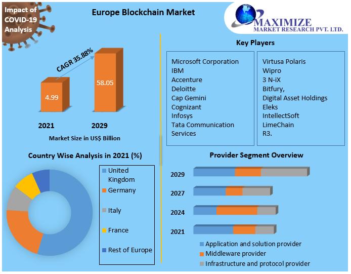
All 5 Releases
More Releases for Tissue
- Categories Advertising, Media Consulting, Marketing Research Arts & Culture Associations & Organizations Business, Economy, Finances, Banking & Insurance Energy & Environment Fashion, Lifestyle, Trends Health & Medicine Industry, Real Estate & Construction IT, New Media & Software Leisure, Entertainment, Miscellaneous Logistics & Transport Media & Telecommunications Politics, Law & Society Science & Education Sports Tourism, Cars, Traffic RSS-Newsfeeds
- Order Credits
- About Us About / FAQ Newsletter Terms & Conditions Privacy Policy Imprint
Information
- Author Services
Initiatives
You are accessing a machine-readable page. In order to be human-readable, please install an RSS reader.
All articles published by MDPI are made immediately available worldwide under an open access license. No special permission is required to reuse all or part of the article published by MDPI, including figures and tables. For articles published under an open access Creative Common CC BY license, any part of the article may be reused without permission provided that the original article is clearly cited. For more information, please refer to https://www.mdpi.com/openaccess .
Feature papers represent the most advanced research with significant potential for high impact in the field. A Feature Paper should be a substantial original Article that involves several techniques or approaches, provides an outlook for future research directions and describes possible research applications.
Feature papers are submitted upon individual invitation or recommendation by the scientific editors and must receive positive feedback from the reviewers.
Editor’s Choice articles are based on recommendations by the scientific editors of MDPI journals from around the world. Editors select a small number of articles recently published in the journal that they believe will be particularly interesting to readers, or important in the respective research area. The aim is to provide a snapshot of some of the most exciting work published in the various research areas of the journal.
Original Submission Date Received: .
- Active Journals
- Find a Journal
- Proceedings Series
- For Authors
- For Reviewers
- For Editors
- For Librarians
- For Publishers
- For Societies
- For Conference Organizers
- Open Access Policy
- Institutional Open Access Program
- Special Issues Guidelines
- Editorial Process
- Research and Publication Ethics
- Article Processing Charges
- Testimonials
- Preprints.org
- SciProfiles
- Encyclopedia
Article Menu

- Subscribe SciFeed
- Recommended Articles
- Google Scholar
- on Google Scholar
- Table of Contents
Find support for a specific problem in the support section of our website.
Please let us know what you think of our products and services.
Visit our dedicated information section to learn more about MDPI.
JSmol Viewer
Regenerative cosmetics: skin tissue engineering for anti-aging, repair, and hair restoration.

1. Introduction
1.1. importance of healthy skin and hair in society, 1.2. limitations of traditional cosmetic approaches, 1.3. the promise of te-based dermocosmetics.
- Addressing the root cause: TE-based dermocosmetics aim to address the underlying biological processes responsible for various skin and hair concerns. This can involve stimulating collagen production for anti-aging effects, promoting wound healing through the delivery of growth factors, or even facilitating hair follicle (HF) regeneration through the use of bioengineered scaffolds [ 18 ].
- Enhanced efficacy: By targeting specifically the dermal compartment, dermocosmetics derived from TE, including new delivery methods, improve the efficacy of the bioactive compounds or key proteins such as collagen. This can significantly improve areas like scar regeneration and wound healing [ 19 , 20 ].
- Long-lasting results: Some TE techniques, like the application of stem cells or their exosomes, show promise for promoting long-lasting results by stimulating cell proliferation and collagen production. This can significantly reduce the need for frequent product application and improve patient compliance [ 21 , 22 ].
- Better testers: Ex vivo skin models, such as 3D “skin-on-a-chip” (SoC) systems combined with microfluidics, offer a promising alternative to traditional testing methods. These models provide a more realistic recreation of human skin architecture and function, enabling more accurate dermocosmetic product testing [ 23 , 24 ].
2. Mechanisms Involved in Regenerative Cosmetics
2.1. skin aging.
- Skin aging is characterized by a decline in collagen production and a reduction of cell proliferation, together with a decrease in stemness from each tissue, among other factors [ 25 ]. Regenerative cosmetics offer solutions to combat these age-related changes, here is a summary of the key approaches in regenerative medicine applied to address age-related skin changes ( Table 2 ).
2.2. Oxidative Stress
2.3. repair vs. regeneration.
- Promoting wound healing: Engineered skin substitutes like biocompatible scaffolds provide a structure for cell migration and tissue regeneration, accelerating wound healing and minimizing scar formation [ 53 ]. Current skin substitutes have been tested for addressing regeneration in burn patients, chronic ulcers (diabetes), and rare genodermatoses (Epidermolysis bullosa) [ 54 ]. Novel technologies, including injectable cell suspensions and 3D scaffolds, are promising for improving wound healing and skin regeneration [ 55 ].
- Scar reduction: Microneedling and fractional laser therapy, combined with regenerative ingredients like growth factors, can stimulate collagen production and improve the appearance of existing scars [ 56 , 57 ]. Additionally, platelet-rich plasma (PRP) therapy is gaining traction as a potential scar reduction technique. Studies suggest that PRP injections may improve scar quality and reduce scar tissue formation [ 58 ], which is particularly interesting in relation to acne scars.
2.4. Fibrosis and Connective Tissue in Skin Rejuvenation
- Modulate the fibrotic process: by understanding the molecular mechanisms underlying fibrosis, researchers can develop strategies to control collagen deposition and promote scarless wound healing, and also highlight the role of macrophages in the inflammatory phase [ 59 ].
- Enhance the functionality of the connective tissue: supporting the health and organization of the connective tissue, which provides structural support and elasticity to the skin, is crucial for maintaining a youthful appearance and function, and this is particularly interesting when the role of MSCs is studied in UV-associated skin aging [ 61 ].
2.5. Hair Follicle Regeneration
3. regenerative cosmetics: a transformative alternative, 3.1. omic approaches: a key tool for regenerative cosmetics, 3.1.1. proteomics, 3.1.2. metabolomics, 3.1.3. multi-omics integration, 3.2. skin modeling accelerates drug development, 3d skin-on-chip models and microfluidics for dermocosmetics.
- Materials: The 3D SoC models require balancing cost and biomimicry through material selection. Synthetic polymers (PDMS, PCL, PLA) offer affordability and biocompatibility but lack the intricate structure of natural tissues. Hydrogels (alginate, collagen) mimic tissues but struggle with maintaining precise mechanical properties. Animal-derived materials (decellularized ECM, silk) provide the most biomimetic environment, closely resembling natural skin, but are expensive and raise ethical concerns [ 95 , 98 , 99 , 100 , 101 , 102 ]. Recent innovations like decellularized ECM and silk offer promising solutions, aiming to bridge the gap between affordability and biomimicry [ 15 , 103 ].
- Challenges : SoC models face challenges in controlling chemical gradients, technical sampling, and analysis. Integrating vasculature and microbiomes is crucial for physiological accuracy. Despite these, models like EpiDerm from MatTek Corporation or SkinEthic from L’Oréal show promise for dermocosmetics, offering safety and efficacy benefits over traditional methods [ 93 , 104 , 105 ].
3.3. Potential Solutions for Hair Loss and Promotion of Thicker, Healthier Hair Growth
- Low-level laser therapy (LLLT) stimulates hair growth with minimal side effects by exposing tissues to low-level light energy, showing a synergistic effect on promoting hair regrowth [ 107 ].
- Mesotherapy , involving the intradermal infusion of a mixture of factors, has demonstrated improvements in hair growth [ 93 , 108 , 109 ].
- Carboxytherapy , which entails the intradermic or subcutaneous insufflation of medical-grade sterile CO 2 , enhances blood flow and nutrient delivery to the HF, potentially aiding in cases of alopecia [ 93 , 110 , 111 ].
- Microneedling promotes hair growth by inducing percutaneous wounds with sterile microneedles, stimulating the activation of HFSCs and the release of growth factors that promote wound healing and angiogenesis [ 112 , 113 ].
- Autologous PRP treatment, derived from the patient’s blood, stimulates hair growth through the release of growth factors, cytokines, and chemokines, promoting cell proliferation, differentiation, and angiogenesis [ 114 ].
- Nanoparticles have been studied for drug delivery directly into the HF, minimizing the systemic adverse effects [ 107 ].
4. Revolutionizing Beauty: The Convergence of Regenerative Medicine and Cosmetic Science
5. challenges, future opportunities, and the role of ai in this field, 6. conclusions, author contributions, institutional review board statement, informed consent statement, data availability statement, acknowledgments, conflicts of interest.
- Bouwstra, J.A.; Nadaban, A.; Bras, W.; McCabe, C.; Bunge, A.; Gooris, G.S. The skin barrier: An extraordinary interface with an exceptional lipid organization. Prog. Lipid Res. 2023 , 92 , 101252. [ Google Scholar ] [ CrossRef ]
- Mansbridge, J. Skin tissue engineering. J. Biomater. Sci. Polym. Ed. 2008 , 19 , 955–968. [ Google Scholar ] [ CrossRef ] [ PubMed ]
- Lasisi, T.; Smallcombe, J.W.; Kenney, W.L.; Shriver, M.D.; Zydney, B.; Jablonski, N.G.; Havenith, G. Human scalp hair as a thermoregulatory adaptation. Proc. Natl. Acad. Sci. USA 2023 , 120 , e2301760120. [ Google Scholar ] [ CrossRef ] [ PubMed ]
- Silverberg, J.I. Comorbidities and the impact of atopic dermatitis. Ann. Allergy Asthma Immunol. 2019 , 123 , 144–151. [ Google Scholar ] [ CrossRef ]
- Hwang, H.W.; Ryou, S.; Jeong, J.H.; Lee, J.W.; Lee, K.J.; Lee, S.B.; Shin, H.T.; Byun, J.W.; Shin, J.; Choi, G.S. The Quality of Life and Psychosocial Impact on Female Pattern Hair Loss. Ann. Dermatol. 2024 , 36 , 44–52. [ Google Scholar ] [ CrossRef ] [ PubMed ]
- Ahluwalia, J.; Fabi, S.G. The psychological and aesthetic impact of age-related hair changes in females. J. Cosmet. Dermatol. 2019 , 18 , 1161–1169. [ Google Scholar ] [ CrossRef ]
- Schielein, M.C.; Tizek, L.; Ziehfreund, S.; Sommer, R.; Biedermann, T.; Zink, A. Stigmatization caused by hair loss—A systematic literature review. JDDG J. Der Dtsch. Dermatol. Ges. 2020 , 18 , 1357–1368. [ Google Scholar ] [ CrossRef ]
- Nandy, P.; Shrivastava, T. Exploring the Multifaceted Impact of Acne on Quality of Life and Well-Being. Cureus 2024 , 16 , e52727. [ Google Scholar ] [ CrossRef ]
- Bertoli, M.J.; Sadoughifar, R.; Schwartz, R.A.; Lotti, T.M.; Janniger, C.K. Female pattern hair loss: A comprehensive review. Dermatol. Ther. 2020 , 33 , e14055. [ Google Scholar ] [ CrossRef ]
- York, K.; Meah, N.; Bhoyrul, B.; Sinclair, R. A review of the treatment of male pattern hair loss. Expert Opin. Pharmacother. 2020 , 21 , 603–612. [ Google Scholar ] [ CrossRef ]
- Dhapte-Pawar, V.; Kadam, S.; Saptarsi, S.; Kenjale, P.P. Nanocosmeceuticals: Facets and aspects. Future Sci. OA 2020 , 6 , FSO613. [ Google Scholar ] [ CrossRef ] [ PubMed ]
- Vyas, K.S.; Kaufman, J.; Munavalli, G.S.; Robertson, K.; Behfar, A.; Wyles, S.P. Exosomes: The latest in regenerative aesthetics. Regen. Med. 2023 , 18 , 181–194. [ Google Scholar ] [ CrossRef ] [ PubMed ]
- Mathes, S.H.; Ruffner, H.; Graf-Hausner, U. The use of skin models in drug development. Adv. Drug Deliv. Rev. 2014 , 69–70 , 81–102. [ Google Scholar ] [ CrossRef ] [ PubMed ]
- Sarkiri, M.; Fox, S.C.; Fratila-Apachitei, L.E.; Zadpoor, A.A. Bioengineered Skin Intended for Skin Disease Modeling. Int. J. Mol. Sci. 2019 , 20 , 1407. [ Google Scholar ] [ CrossRef ] [ PubMed ]
- Gao, G.; Yonezawa, T.; Hubbell, K.; Dai, G.; Cui, X. Inkjet-bioprinted acrylated peptides and PEG hydrogel with human mesenchymal stem cells promote robust bone and cartilage formation with minimal printhead clogging. Biotechnol. J. 2015 , 10 , 1568–1577. [ Google Scholar ] [ CrossRef ] [ PubMed ]
- Yin, J.; Yan, M.; Wang, Y.; Fu, J.; Suo, H. 3D Bioprinting of Low-Concentration Cell-Laden Gelatin Methacrylate (GelMA) Bioinks with a Two-Step Cross-linking Strategy. ACS Appl. Mater. Interfaces 2018 , 10 , 6849–6857. [ Google Scholar ] [ CrossRef ] [ PubMed ]
- Jin, Z.; Li, Y.; Yu, K.; Liu, L.; Fu, J.; Yao, X.; Zhang, A.; He, Y. 3D Printing of Physical Organ Models: Recent Developments and Challenges. Adv. Sci. 2021 , 8 , e2101394. [ Google Scholar ] [ CrossRef ] [ PubMed ]
- Khalili, M.H.; Zhang, R.; Wilson, S.; Goel, S.; Impey, S.A.; Aria, A.I. Additive Manufacturing and Physicomechanical Characteristics of PEGDA Hydrogels: Recent Advances and Perspective for Tissue Engineering. Polymers 2023 , 15 , 2341. [ Google Scholar ] [ CrossRef ] [ PubMed ]
- Matama, T.; Costa, C.; Fernandes, B.; Araujo, R.; Cruz, C.F.; Tortosa, F.; Sheeba, C.J.; Becker, J.D.; Gomes, A.; Cavaco-Paulo, A. Changing human hair fibre colour and shape from the follicle. J. Adv. Res. 2023 . [ Google Scholar ] [ CrossRef ]
- Monavarian, M.; Kader, S.; Moeinzadeh, S.; Jabbari, E. Regenerative Scar-Free Skin Wound Healing. Tissue Eng. Part B Rev. 2019 , 25 , 294–311. [ Google Scholar ] [ CrossRef ]
- Dehkordi, A.N.; Babaheydari, F.M.; Chehelgerdi, M.; Dehkordi, S.R. Skin tissue engineering: Wound healing based on stem-cell-based therapeutic strategies. Stem Cell Res. Ther. 2019 , 10 , 111. [ Google Scholar ] [ CrossRef ] [ PubMed ]
- Jo, H.; Brito, S.; Kwak, B.M.; Park, S.; Lee, M.G.; Bin, B.H. Applications of Mesenchymal Stem Cells in Skin Regeneration and Rejuvenation. Int. J. Mol. Sci. 2021 , 22 , 2410. [ Google Scholar ] [ CrossRef ] [ PubMed ]
- Zhou, C.; Zhang, B.; Yang, Y.; Jiang, Q.; Li, T.; Gong, J.; Tang, H.; Zhang, Q. Stem cell-derived exosomes: Emerging therapeutic opportunities for wound healing. Stem Cell Res. Ther. 2023 , 14 , 107. [ Google Scholar ] [ CrossRef ] [ PubMed ]
- Eberlin, S.; Silva, M.S.D.; Facchini, G.; Silva, G.H.D.; Pinheiro, A.; Eberlin, S.; Pinheiro, A.D.S. The Ex Vivo Skin Model as an Alternative Tool for the Efficacy and Safety Evaluation of Topical Products. Altern. Lab. Anim. 2020 , 48 , 10–22. [ Google Scholar ] [ CrossRef ] [ PubMed ]
- Bataillon, M.; Lelievre, D.; Chapuis, A.; Thillou, F.; Autourde, J.B.; Durand, S.; Boyera, N.; Rigaudeau, A.S.; Besne, I.; Pellevoisin, C. Characterization of a New Reconstructed Full Thickness Skin Model, T-Skin, and its Application for Investigations of Anti-Aging Compounds. Int. J. Mol. Sci. 2019 , 20 , 2240. [ Google Scholar ] [ CrossRef ]
- Shin, S.H.; Lee, Y.H.; Rho, N.K.; Park, K.Y. Skin aging from mechanisms to interventions: Focusing on dermal aging. Front. Physiol. 2023 , 14 , 1195272. [ Google Scholar ] [ CrossRef ]
- Gref, R.; Delomenie, C.; Maksimenko, A.; Gouadon, E.; Percoco, G.; Lati, E.; Desmaele, D.; Zouhiri, F.; Couvreur, P. Vitamin C-squalene bioconjugate promotes epidermal thickening and collagen production in human skin. Sci. Rep. 2020 , 10 , 16883. [ Google Scholar ] [ CrossRef ] [ PubMed ]
- Skibska, A.; Perlikowska, R. Signal Peptides—Promising Ingredients in Cosmetics. Curr. Protein Pept. Sci. 2021 , 22 , 716–728. [ Google Scholar ] [ CrossRef ]
- Bukhari, S.N.A.; Roswandi, N.L.; Waqas, M.; Habib, H.; Hussain, F.; Khan, S.; Sohail, M.; Ramli, N.A.; Thu, H.E.; Hussain, Z. Hyaluronic acid, a promising skin rejuvenating biomedicine: A review of recent updates and pre-clinical and clinical investigations on cosmetic and nutricosmetic effects. Int. J. Biol. Macromol. 2018 , 120 , 1682–1695. [ Google Scholar ] [ CrossRef ]
- Spataro, E.A.; Dierks, K.; Carniol, P.J. Microneedling-Associated Procedures to Enhance Facial Rejuvenation. Facial Plast. Surg. Clin. N. Am. 2022 , 30 , 389–397. [ Google Scholar ] [ CrossRef ]
- Kim, Y.J.; Yoo, S.M.; Park, H.H.; Lim, H.J.; Kim, Y.L.; Lee, S.; Seo, K.W.; Kang, K.S. Exosomes derived from human umbilical cord blood mesenchymal stem cells stimulates rejuvenation of human skin. Biochem. Biophys. Res. Commun. 2017 , 493 , 1102–1108. [ Google Scholar ] [ CrossRef ] [ PubMed ]
- Sierra-Sanchez, A.; Kim, K.H.; Blasco-Morente, G.; Arias-Santiago, S. Cellular human tissue-engineered skin substitutes investigated for deep and difficult to heal injuries. NPJ Regen. Med. 2021 , 6 , 35. [ Google Scholar ] [ CrossRef ] [ PubMed ]
- Baek, J.; Lee, M.G. Oxidative stress and antioxidant strategies in dermatology. Redox Rep. 2016 , 21 , 164–169. [ Google Scholar ] [ CrossRef ] [ PubMed ]
- Trouba, K.J.; Hamadeh, H.K.; Amin, R.P.; Germolec, D.R. Oxidative stress and its role in skin disease. Antioxid. Redox Signal. 2002 , 4 , 665–673. [ Google Scholar ] [ CrossRef ] [ PubMed ]
- Popa, G.L.; Mitran, C.I.; Mitran, M.I.; Tampa, M.; Matei, C.; Popa, M.I.; Georgescu, S.R. Markers of Oxidative Stress in Patients with Acne: A Literature Review. Life 2023 , 13 , 1433. [ Google Scholar ] [ CrossRef ] [ PubMed ]
- Jones, D.A. Rosacea, reactive oxygen species, and azelaic Acid. J. Clin. Aesthet. Dermatol. 2009 , 2 , 26–30. [ Google Scholar ] [ PubMed ]
- Rahimi, H.; Mirnezami, M.; Yazdabadi, A.; Hajihashemi, A. Evaluation of systemic oxidative stress in patients with melasma. J. Cosmet. Dermatol. 2024 , 23 , 284–288. [ Google Scholar ] [ CrossRef ] [ PubMed ]
- Prie, B.E.; Voiculescu, V.M.; Ionescu-Bozdog, O.B.; Petrutescu, B.; Iosif, L.; Gaman, L.E.; Clatici, V.G.; Stoian, I.; Giurcaneanu, C. Oxidative stress and alopecia areata. J. Med. Life 2015 , 8 , 43–46. [ Google Scholar ]
- Zapatero-Solana, E.; Garcia-Gimenez, J.L.; Guerrero-Aspizua, S.; Garcia, M.; Toll, A.; Baselga, E.; Duran-Moreno, M.; Markovic, J.; Garcia-Verdugo, J.M.; Conti, C.J.; et al. Oxidative stress and mitochondrial dysfunction in Kindler syndrome. Orphanet J. Rare Dis. 2014 , 9 , 211. [ Google Scholar ] [ CrossRef ]
- Padayatty, S.J.; Katz, A.; Wang, Y.; Eck, P.; Kwon, O.; Lee, J.H.; Chen, S.; Corpe, C.; Dutta, A.; Dutta, S.K.; et al. Vitamin C as an antioxidant: Evaluation of its role in disease prevention. J. Am. Coll. Nutr. 2003 , 22 , 18–35. [ Google Scholar ] [ CrossRef ]
- Niki, E. Interaction of ascorbate and alpha-tocopherol. Ann. N. Y. Acad. Sci. 1987 , 498 , 186–199. [ Google Scholar ] [ CrossRef ] [ PubMed ]
- Farris, P.K. Topical vitamin C: A useful agent for treating photoaging and other dermatologic conditions. Dermatol. Surg. 2005 , 31 , 814–817; discussion 818. [ Google Scholar ] [ CrossRef ] [ PubMed ]
- Turcov, D.; Zbranca-Toporas, A.; Suteu, D. Bioactive Compounds for Combating Oxidative Stress in Dermatology. Int. J. Mol. Sci. 2023 , 24 , 17517. [ Google Scholar ] [ CrossRef ] [ PubMed ]
- Shaban, S.; El-Husseny, M.W.A.; Abushouk, A.I.; Salem, A.M.A.; Mamdouh, M.; Abdel-Daim, M.M. Effects of Antioxidant Supplements on the Survival and Differentiation of Stem Cells. Oxidative Med. Cell. Longev. 2017 , 2017 , 5032102. [ Google Scholar ] [ CrossRef ]
- Luo, L.; Kawakatsu, M.; Guo, C.W.; Urata, Y.; Huang, W.J.; Ali, H.; Doi, H.; Kitajima, Y.; Tanaka, T.; Goto, S.; et al. Effects of antioxidants on the quality and genomic stability of induced pluripotent stem cells. Sci. Rep. 2014 , 4 , 3779. [ Google Scholar ] [ CrossRef ] [ PubMed ]
- Choi, K.M.; Seo, Y.K.; Yoon, H.H.; Song, K.Y.; Kwon, S.Y.; Lee, H.S.; Park, J.K. Effect of ascorbic acid on bone marrow-derived mesenchymal stem cell proliferation and differentiation. J. Biosci. Bioeng. 2008 , 105 , 586–594. [ Google Scholar ] [ CrossRef ] [ PubMed ]
- Sies, H.; Jones, D.P. Reactive oxygen species (ROS) as pleiotropic physiological signalling agents. Nat. Rev. Mol. Cell Biol. 2020 , 21 , 363–383. [ Google Scholar ] [ CrossRef ] [ PubMed ]
- Loo, A.E.; Wong, Y.T.; Ho, R.; Wasser, M.; Du, T.; Ng, W.T.; Halliwell, B. Effects of hydrogen peroxide on wound healing in mice in relation to oxidative damage. PLoS ONE 2012 , 7 , e49215. [ Google Scholar ] [ CrossRef ]
- Lyublinskaya, O.G.; Borisov, Y.G.; Pugovkina, N.A.; Smirnova, I.S.; Obidina, J.V.; Ivanova, J.S.; Zenin, V.V.; Shatrova, A.N.; Borodkina, A.V.; Aksenov, N.D.; et al. Reactive Oxygen Species Are Required for Human Mesenchymal Stem Cells to Initiate Proliferation after the Quiescence Exit. Oxidative Med. Cell. Longev. 2015 , 2015 , 502105. [ Google Scholar ] [ CrossRef ]
- De Deken, X.; Corvilain, B.; Dumont, J.E.; Miot, F. Roles of DUOX-mediated hydrogen peroxide in metabolism, host defense, and signaling. Antioxid. Redox Signal. 2014 , 20 , 2776–2793. [ Google Scholar ] [ CrossRef ]
- Guo, L.; Du, J.; Yuan, D.F.; Zhang, Y.; Zhang, S.; Zhang, H.C.; Mi, J.W.; Ning, Y.L.; Chen, M.J.; Wen, D.L.; et al. Optimal H 2 O 2 preconditioning to improve bone marrow mesenchymal stem cells’ engraftment in wound healing. Stem Cell Res. Ther. 2020 , 11 , 434. [ Google Scholar ] [ CrossRef ]
- Buron, M.; Palomares, T.; Garrido-Pascual, P.; Herrero de la Parte, B.; Garcia-Alonso, I.; Alonso-Varona, A. Conditioned Medium from H 2 O 2 —Preconditioned Human Adipose-Derived Stem Cells Ameliorates UVB-Induced Damage to Human Dermal Fibroblasts. Antioxidants 2022 , 11 , 2011. [ Google Scholar ] [ CrossRef ]
- Lee, Y.; Son, J.Y.; Kang, J.I.; Park, K.M.; Park, K.D. Hydrogen Peroxide-Releasing Hydrogels for Enhanced Endothelial Cell Activities and Neovascularization. ACS Appl. Mater. Interfaces 2018 , 10 , 18372–18379. [ Google Scholar ] [ CrossRef ]
- Pleguezuelos-Beltran, P.; Galvez-Martin, P.; Nieto-Garcia, D.; Marchal, J.A.; Lopez-Ruiz, E. Advances in spray products for skin regeneration. Bioact. Mater. 2022 , 16 , 187–203. [ Google Scholar ] [ CrossRef ]
- Martinez-Santamaria, L.; Guerrero-Aspizua, S.; Del Rio, M. Skin bioengineering: Preclinical and clinical applications. Actas Dermosifiliogr. 2012 , 103 , 5–11. [ Google Scholar ] [ CrossRef ]
- Chocarro-Wrona, C.; Lopez-Ruiz, E.; Peran, M.; Galvez-Martin, P.; Marchal, J.A. Therapeutic strategies for skin regeneration based on biomedical substitutes. J. Eur. Acad. Dermatol. Venereol. 2019 , 33 , 484–496. [ Google Scholar ] [ CrossRef ]
- Disphanurat, W.; Sivapornpan, N.; Srisantithum, B.; Leelawattanachai, J. Efficacy of a triamcinolone acetonide-loaded dissolving microneedle patch for the treatment of hypertrophic scars and keloids: A randomized, double-blinded, placebo-controlled split-scar study. Arch. Dermatol. Res. 2023 , 315 , 989–997. [ Google Scholar ] [ CrossRef ]
- Waghmare, K.B.; Sequeira, J.; Rao, B.H.S. An objective assessment of microneedling therapy in atrophic facial acne scars. Natl. J. Maxillofac. Surg. 2022 , 13 , S103–S107. [ Google Scholar ] [ CrossRef ]
- Long, T.; Gupta, A.; Ma, S.; Hsu, S. Platelet-rich plasma in noninvasive procedures for atrophic acne scars: A systematic review and meta-analysis. J. Cosmet. Dermatol. 2020 , 19 , 836–844. [ Google Scholar ] [ CrossRef ]
- Mony, M.P.; Harmon, K.A.; Hess, R.; Dorafshar, A.H.; Shafikhani, S.H. An Updated Review of Hypertrophic Scarring. Cells 2023 , 12 , 678. [ Google Scholar ] [ CrossRef ]
- Tartaglia, G.; Cao, Q.; Padron, Z.M.; South, A.P. Impaired Wound Healing, Fibrosis, and Cancer: The Paradigm of Recessive Dystrophic Epidermolysis Bullosa. Int. J. Mol. Sci. 2021 , 22 , 5104. [ Google Scholar ] [ CrossRef ]
- Cao, Z.; Jin, S.; Wang, P.; He, Q.; Yang, Y.; Gao, Z.; Wang, X. Microneedle based adipose derived stem cells-derived extracellular vesicles therapy ameliorates UV-induced photoaging in SKH-1 mice. J. Biomed. Mater. Res. A 2021 , 109 , 1849–1857. [ Google Scholar ] [ CrossRef ]
- Sorg, H.; Tilkorn, D.J.; Hager, S.; Hauser, J.; Mirastschijski, U. Skin Wound Healing: An Update on the Current Knowledge and Concepts. Eur. Surg. Res. 2017 , 58 , 81–94. [ Google Scholar ] [ CrossRef ]
- Lin, X.; Zhu, L.; He, J. Morphogenesis, Growth Cycle and Molecular Regulation of Hair Follicles. Front. Cell Dev. Biol. 2022 , 10 , 899095. [ Google Scholar ] [ CrossRef ]
- Vandishi, A.K.; Esmaeili, A.; Taghipour, N. The promising prospect of human hair follicle regeneration in the shadow of new tissue engineering strategies. Tissue Cell 2024 , 87 , 102338. [ Google Scholar ] [ CrossRef ]
- Llamas-Molina, J.M.; Carrero-Castano, A.; Ruiz-Villaverde, R.; Campos, A. Tissue Engineering and Regeneration of the Human Hair Follicle in Androgenetic Alopecia: Literature Review. Life 2022 , 12 , 117. [ Google Scholar ] [ CrossRef ]
- Castro, A.R.; Logarinho, E. Tissue engineering strategies for human hair follicle regeneration: How far from a hairy goal? Stem Cells Transl. Med. 2020 , 9 , 342–350. [ Google Scholar ] [ CrossRef ]
- Xu, K.; Yu, E.; Wu, M.; Wei, P.; Yin, J. Cells, growth factors and biomaterials used in tissue engineering for hair follicles regeneration. Regen. Ther. 2022 , 21 , 596–610. [ Google Scholar ] [ CrossRef ]
- Kageyama, T.; Yan, L.; Shimizu, A.; Maruo, S.; Fukuda, J. Preparation of hair beads and hair follicle germs for regenerative medicine. Biomaterials 2019 , 212 , 55–63. [ Google Scholar ] [ CrossRef ]
- Wang, J.; Miao, Y.; Huang, Y.; Lin, B.; Liu, X.; Xiao, S.; Du, L.; Hu, Z.; Xing, M. Bottom-up Nanoencapsulation from Single Cells to Tunable and Scalable Cellular Spheroids for Hair Follicle Regeneration. Adv. Healthc. Mater. 2018 , 7 , 1700447. [ Google Scholar ] [ CrossRef ]
- Zhang, Y.; Yin, P.; Huang, J.; Yang, L.; Liu, Z.; Fu, D.; Hu, Z.; Huang, W.; Miao, Y. Scalable and high-throughput production of an injectable platelet-rich plasma (PRP)/cell-laden microcarrier/hydrogel composite system for hair follicle tissue engineering. J. Nanobiotechnol. 2022 , 20 , 465. [ Google Scholar ] [ CrossRef ] [ PubMed ]
- Kageyama, T.; Nanmo, A.; Yan, L.; Nittami, T.; Fukuda, J. Effects of platelet-rich plasma on in vitro hair follicle germ preparation for hair regenerative medicine. J. Biosci. Bioeng. 2020 , 130 , 666–671. [ Google Scholar ] [ CrossRef ] [ PubMed ]
- Zhang, K.; Bai, X.; Yuan, Z.; Cao, X.; Jiao, X.; Qin, Y.; Wen, Y.; Zhang, X. Cellular Nanofiber Structure with Secretory Activity-Promoting Characteristics for Multicellular Spheroid Formation and Hair Follicle Regeneration. ACS Appl. Mater. Interfaces 2020 , 12 , 7931–7941. [ Google Scholar ] [ CrossRef ] [ PubMed ]
- Fernandez-Martos, S.; Calvo-Sanchez, M.; Garcia-Alonso, K.; Castro, B.; Hashtroody, B.; Espada, J. Sustained Human Hair Follicle Growth Ex Vivo in a Glycosaminoglycan Hydrogel Matrix. Int. J. Mol. Sci. 2019 , 20 , 1741. [ Google Scholar ] [ CrossRef ] [ PubMed ]
- Xie, S.; Chen, L.; Zhang, M.; Zhang, C.; Li, H. Self-assembled complete hair follicle organoids by coculture of neonatal mouse epidermal cells and dermal cells in Matrigel. Ann. Transl. Med. 2022 , 10 , 767. [ Google Scholar ] [ CrossRef ] [ PubMed ]
- Zhang, X.; Xiao, S.; Liu, B.; Miao, Y.; Hu, Z. Use of extracellular matrix hydrogel from human placenta to restore hair-inductive potential of dermal papilla cells. Regen. Med. 2019 , 14 , 741–751. [ Google Scholar ] [ CrossRef ] [ PubMed ]
- Barat, T.; Abdollahimajd, F.; Dadkhahfar, S.; Moravvej, H. Evaluation of the efficacy and safety of cow placenta extract lotion versus minoxidil 2% in the treatment of female pattern androgenetic alopecia. Int. J. Women’s Dermatol. 2020 , 6 , 318–321. [ Google Scholar ] [ CrossRef ] [ PubMed ]
- Motter Catarino, C.; Cigaran Schuck, D.; Dechiario, L.; Karande, P. Incorporation of hair follicles in 3D bioprinted models of human skin. Sci. Adv. 2023 , 9 , eadg0297. [ Google Scholar ] [ CrossRef ]
- Kang, D.; Liu, Z.; Qian, C.; Huang, J.; Zhou, Y.; Mao, X.; Qu, Q.; Liu, B.; Wang, J.; Hu, Z.; et al. 3D bioprinting of a gelatin-alginate hydrogel for tissue-engineered hair follicle regeneration. Acta Biomater. 2023 , 165 , 19–30. [ Google Scholar ] [ CrossRef ]
- Kang, M.S.; Kwon, M.; Lee, S.H.; Kim, W.H.; Lee, G.W.; Jo, H.J.; Kim, B.; Yang, S.Y.; Kim, K.S.; Han, D.W. 3D Printing of Skin Equivalents with Hair Follicle Structures and Epidermal-Papillary-Dermal Layers Using Gelatin/Hyaluronic Acid Hydrogels. Chem. Asian J. 2022 , 17 , e202200620. [ Google Scholar ] [ CrossRef ]
- Zheng, S.Y.; Hu, X.M.; Huang, K.; Li, Z.H.; Chen, Q.N.; Yang, R.H.; Xiong, K. Proteomics as a tool to improve novel insights into skin diseases: What we know and where we should be going. Front. Surg. 2022 , 9 , 1025557. [ Google Scholar ] [ CrossRef ] [ PubMed ]
- Benoit, I.; Burty-Valin, E.; Radman, M. A Proteome-Centric View of Ageing, including that of the Skin and Age-Related Diseases: Considerations of a Common Cause and Common Preventative and Curative Interventions. Clin. Cosmet. Investig. Dermatol. 2023 , 16 , 79–85. [ Google Scholar ] [ CrossRef ] [ PubMed ]
- Pien, N.; Bray, F.; Gheysens, T.; Tytgat, L.; Rolando, C.; Mantovani, D.; Dubruel, P.; Vlierberghe, S.V. Proteomics as a tool to gain next level insights into photo-crosslinkable biopolymer modifications. Bioact. Mater. 2022 , 17 , 204–220. [ Google Scholar ] [ CrossRef ] [ PubMed ]
- Tan, K.X.; Chang, T.; Lin, X. Secretomes as an emerging class of bioactive ingredients for enhanced cosmeceutical applications. Exp. Dermatol. 2022 , 31 , 674–688. [ Google Scholar ] [ CrossRef ] [ PubMed ]
- Masutin, V.; Kersch, C.; Schmitz-Spanke, S. A systematic review: Metabolomics-based identification of altered metabolites and pathways in the skin caused by internal and external factors. Exp. Dermatol. 2022 , 31 , 700–714. [ Google Scholar ] [ CrossRef ]
- Knox, S.; O’Boyle, N.M. Skin lipids in health and disease: A review. Chem. Phys. Lipids 2021 , 236 , 105055. [ Google Scholar ] [ CrossRef ] [ PubMed ]
- Chen, T.; Zhu, Z.; Du, Q.; Wang, Z.; Wu, W.; Xue, Y.; Wang, Y.; Wu, Y.; Zeng, Q.; Jiang, C.; et al. A Skin Lipidomics Study Reveals the Therapeutic Effects of Tanshinones in a Rat Model of Acne. Front. Pharmacol. 2021 , 12 , 675659. [ Google Scholar ] [ CrossRef ] [ PubMed ]
- Chen, H.; Zhao, Q.; Zhong, Q.; Duan, C.; Krutmann, J.; Wang, J.; Xia, J. Skin Microbiome, Metabolome and Skin Phenome, from the Perspectives of Skin as an Ecosystem. Phenomics 2022 , 2 , 363–382. [ Google Scholar ] [ CrossRef ] [ PubMed ]
- Gueniche, A.; Perin, O.; Bouslimani, A.; Landemaine, L.; Misra, N.; Cupferman, S.; Aguilar, L.; Clavaud, C.; Chopra, T.; Khodr, A. Advances in Microbiome-Derived Solutions and Methodologies Are Founding a New Era in Skin Health and Care. Pathogens 2022 , 11 , 121. [ Google Scholar ] [ CrossRef ]
- Tong, T.; Geng, R.; Kang, S.G.; Li, X.; Huang, K. Revitalizing Photoaging Skin through Eugenol in UVB-Exposed Hairless Mice: Mechanistic Insights from Integrated Multi-Omics. Antioxidants 2024 , 13 , 168. [ Google Scholar ] [ CrossRef ]
- Bonafont, J.; Mencia, A.; Garcia, M.; Torres, R.; Rodriguez, S.; Carretero, M.; Chacon-Solano, E.; Modamio-Hoybjor, S.; Marinas, L.; Leon, C.; et al. Clinically Relevant Correction of Recessive Dystrophic Epidermolysis Bullosa by Dual sgRNA CRISPR/Cas9-Mediated Gene Editing. Mol. Ther. 2019 , 27 , 986–998. [ Google Scholar ] [ CrossRef ]
- Carretero, M.; Guerrero-Aspizua, S.; Del Rio, M. Bioengineered skin humanized model of psoriasis. Methods Mol. Biol. 2013 , 961 , 305–323. [ Google Scholar ] [ CrossRef ]
- Guerrero-Aspizua, S.; Carretero, M.; Conti, C.J.; Del Rio, M. The importance of immunity in the development of reliable animal models for psoriasis and atopic dermatitis. Immunol. Cell Biol. 2020 , 98 , 626–638. [ Google Scholar ] [ CrossRef ]
- Fernandez-Carro, E.; Angenent, M.; Gracia-Cazana, T.; Gilaberte, Y.; Alcaine, C.; Ciriza, J. Modeling an Optimal 3D Skin-on-Chip within Microfluidic Devices for Pharmacological Studies. Pharmaceutics 2022 , 14 , 1417. [ Google Scholar ] [ CrossRef ]
- Rodrigues, R.O.; Sousa, P.C.; Gaspar, J.; Banobre-Lopez, M.; Lima, R.; Minas, G. Organ-on-a-Chip: A Preclinical Microfluidic Platform for the Progress of Nanomedicine. Small 2020 , 16 , e2003517. [ Google Scholar ] [ CrossRef ]
- Ponmozhi, J.; Dhinakaran, S.; Varga-Medveczky, Z.; Fonagy, K.; Bors, L.A.; Ivan, K.; Erdo, F. Development of Skin-On-A-Chip Platforms for Different Utilizations: Factors to Be Considered. Micromachines 2021 , 12 , 294. [ Google Scholar ] [ CrossRef ]
- Mohamadali, M.; Ghiaseddin, A.; Irani, S.; Amirkhani, M.A.; Dahmardehei, M. Design and evaluation of a skin-on-a-chip pumpless microfluidic device. Sci. Rep. 2023 , 13 , 8861. [ Google Scholar ] [ CrossRef ]
- Wufuer, M.; Lee, G.; Hur, W.; Jeon, B.; Kim, B.J.; Choi, T.H.; Lee, S. Skin-on-a-chip model simulating inflammation, edema and drug-based treatment. Sci. Rep. 2016 , 6 , 37471. [ Google Scholar ] [ CrossRef ]
- Randall, M.J.; Jungel, A.; Rimann, M.; Wuertz-Kozak, K. Advances in the Biofabrication of 3D Skin in vitro: Healthy and Pathological Models. Front. Bioeng. Biotechnol. 2018 , 6 , 154. [ Google Scholar ] [ CrossRef ]
- Bellas, E.; Seiberg, M.; Garlick, J.; Kaplan, D.L. In vitro 3D full-thickness skin-equivalent tissue model using silk and collagen biomaterials. Macromol. Biosci. 2012 , 12 , 1627–1636. [ Google Scholar ] [ CrossRef ]
- Parenteau-Bareil, R.; Gauvin, R.; Cliche, S.; Gariepy, C.; Germain, L.; Berthod, F. Comparative study of bovine, porcine and avian collagens for the production of a tissue engineered dermis. Acta Biomater. 2011 , 7 , 3757–3765. [ Google Scholar ] [ CrossRef ]
- Rosa, E.; Diaferia, C.; Gianolio, E.; Sibillano, T.; Gallo, E.; Smaldone, G.; Stornaiuolo, M.; Giannini, C.; Morelli, G.; Accardo, A. Multicomponent Hydrogel Matrices of Fmoc-FF and Cationic Peptides for Application in Tissue Engineering. Macromol. Biosci. 2022 , 22 , e2200128. [ Google Scholar ] [ CrossRef ]
- Arab, W.T.; Susapto, H.H.; Alhattab, D.; Hauser, C.A.E. Peptide nanogels as a scaffold for fabricating dermal grafts and 3D vascularized skin models. J. Tissue Eng. 2022 , 13 , 20417314221111868. [ Google Scholar ] [ CrossRef ]
- Bal-Ozturk, A.; Miccoli, B.; Avci-Adali, M.; Mogtader, F.; Sharifi, F.; Cecen, B.; Yasayan, G.; Braeken, D.; Alarcin, E. Current Strategies and Future Perspectives of Skin-on-a-Chip Platforms: Innovations, Technical Challenges and Commercial Outlook. Curr. Pharm. Des. 2018 , 24 , 5437–5457. [ Google Scholar ] [ CrossRef ]
- Vurat, M.T.; Ergun, C.; Elcin, A.E.; Elcin, Y.M. 3D Bioprinting of Tissue Models with Customized Bioinks. Adv. Exp. Med. Biol. 2020 , 1249 , 67–84. [ Google Scholar ] [ CrossRef ]
- Jimenez, F.; Alam, M.; Vogel, J.E.; Avram, M. Hair transplantation: Basic overview. J. Am. Acad. Dermatol. 2021 , 85 , 803–814. [ Google Scholar ] [ CrossRef ]
- Katzer, T.; Leite Junior, A.; Beck, R.; da Silva, C. Physiopathology and current treatments of androgenetic alopecia: Going beyond androgens and anti-androgens. Dermatol. Ther. 2019 , 32 , e13059. [ Google Scholar ] [ CrossRef ]
- Gupta, A.K.; Polla Ravi, S.; Wang, T.; Talukder, M.; Starace, M.; Piraccini, B.M. Systematic review of mesotherapy: A novel avenue for the treatment of hair loss. J. Dermatol. Treat. 2023 , 34 , 2245084. [ Google Scholar ] [ CrossRef ]
- Tang, Z.; Hu, Y.; Wang, J.; Fan, Z.; Qu, Q.; Miao, Y. Current application of mesotherapy in pattern hair loss: A systematic review. J. Cosmet. Dermatol. 2022 , 21 , 4184–4193. [ Google Scholar ] [ CrossRef ]
- Bagherani, N.; Smoller, B.R.; Tavoosidana, G.; Ghanadan, A.; Wollina, U.; Lotti, T. An overview of the role of carboxytherapy in dermatology. J. Cosmet. Dermatol. 2023 , 22 , 2399–2407. [ Google Scholar ] [ CrossRef ]
- Kroumpouzos, G.; Arora, G.; Kassir, M.; Galadari, H.; Wollina, U.; Lotti, T.; Grabbe, S.; Goldust, M. Carboxytherapy in dermatology. Clin. Dermatol. 2022 , 40 , 305–309. [ Google Scholar ] [ CrossRef ]
- English, R.S., Jr.; Ruiz, S.; DoAmaral, P. Microneedling and Its Use in Hair Loss Disorders: A Systematic Review. Dermatol. Ther (Heidelb) 2022 , 12 , 41–60. [ Google Scholar ] [ CrossRef ]
- Ocampo-Garza, S.S.; Fabbrocini, G.; Ocampo-Candiani, J.; Cinelli, E.; Villani, A. Micro needling: A novel therapeutic approach for androgenetic alopecia, A Review of Literature. Dermatol. Ther. 2020 , 33 , e14267. [ Google Scholar ] [ CrossRef ]
- Paichitrojjana, A.; Paichitrojjana, A. Platelet Rich Plasma and Its Use in Hair Regrowth: A Review. Drug Des. Dev. Ther. 2022 , 16 , 635–645. [ Google Scholar ] [ CrossRef ]
- Yuan, A.R.; Bian, Q.; Gao, J.Q. Current advances in stem cell-based therapies for hair regeneration. Eur. J. Pharmacol. 2020 , 881 , 173197. [ Google Scholar ] [ CrossRef ]
- Rose, P.T. Advances in Hair Restoration. Dermatol. Clin. 2018 , 36 , 57–62. [ Google Scholar ] [ CrossRef ]
- Kwack, M.H.; Seo, C.H.; Gangadaran, P.; Ahn, B.C.; Kim, M.K.; Kim, J.C.; Sung, Y.K. Exosomes derived from human dermal papilla cells promote hair growth in cultured human hair follicles and augment the hair-inductive capacity of cultured dermal papilla spheres. Exp. Dermatol. 2019 , 28 , 854–857. [ Google Scholar ] [ CrossRef ]
- Mahmoudian-Sani, M.R.; Jamshidi, M.; Asgharzade, S. Combined Growth Factor and Gene Therapy: An Approach for Hair Cell Regeneration and Hearing Recovery. ORL J. Otorhinolaryngol. Relat. Spec. 2018 , 80 , 326–337. [ Google Scholar ] [ CrossRef ] [ PubMed ]
- Elder, A.; Cappelli, M.O.; Ring, C.; Saedi, N. Artificial intelligence in cosmetic dermatology: An update on current trends. Clin. Dermatol. 2024 , 42 , 216–220. [ Google Scholar ] [ CrossRef ]
- Waddell, S.J.; de Andres, M.C.; Tsimbouri, P.M.; Alakpa, E.V.; Cusack, M.; Dalby, M.J.; Oreffo, R.O. Biomimetic oyster shell-replicated topography alters the behaviour of human skeletal stem cells. J. Tissue Eng. 2018 , 9 , 2041731418794007. [ Google Scholar ] [ CrossRef ]
Click here to enlarge figure
| Component | Description | References |
|---|---|---|
| : : | [ , ] | |
| : : : | [ , ] | |
| : | [ , ] |
| Approach | Components | Function | References |
|---|---|---|---|
| (e.g., TGF-β, PDGF, IGF-1) | Promote collagen synthesis by skin cells | [ , ] | |
| (e.g., vitamin C, vitamin A) | Enhance collagen production and skin cell proliferation | [ ] | |
| (e.g., Palmitoyl pentapeptide-4) | Stimulate collagen and elastin synthesis | [ ] | |
| Plump up the skin and reduce the appearance of wrinkles | [ ] | ||
| Create controlled micro-injuries to stimulate collagen production and reduce wrinkle depth | [ ] | ||
| Promote collagen synthesis and improve skin elasticity through paracrine effects | [ ] | ||
| Enhance barrier function, hydration, immune response, and wound healing | [ ] |
| The statements, opinions and data contained in all publications are solely those of the individual author(s) and contributor(s) and not of MDPI and/or the editor(s). MDPI and/or the editor(s) disclaim responsibility for any injury to people or property resulting from any ideas, methods, instructions or products referred to in the content. |
Share and Cite
Pleguezuelos-Beltrán, P.; Herráiz-Gil, S.; Martínez-Moreno, D.; Medraño-Fernandez, I.; León, C.; Guerrero-Aspizua, S. Regenerative Cosmetics: Skin Tissue Engineering for Anti-Aging, Repair, and Hair Restoration. Cosmetics 2024 , 11 , 121. https://doi.org/10.3390/cosmetics11040121
Pleguezuelos-Beltrán P, Herráiz-Gil S, Martínez-Moreno D, Medraño-Fernandez I, León C, Guerrero-Aspizua S. Regenerative Cosmetics: Skin Tissue Engineering for Anti-Aging, Repair, and Hair Restoration. Cosmetics . 2024; 11(4):121. https://doi.org/10.3390/cosmetics11040121
Pleguezuelos-Beltrán, Paula, Sara Herráiz-Gil, Daniel Martínez-Moreno, Iria Medraño-Fernandez, Carlos León, and Sara Guerrero-Aspizua. 2024. "Regenerative Cosmetics: Skin Tissue Engineering for Anti-Aging, Repair, and Hair Restoration" Cosmetics 11, no. 4: 121. https://doi.org/10.3390/cosmetics11040121
Article Metrics
Article access statistics, further information, mdpi initiatives, follow mdpi.
Subscribe to receive issue release notifications and newsletters from MDPI journals
IMAGES
VIDEO
COMMENTS
Global paper production has been steadily increasing over the years and reached approximately 425 million tons per year in 2020 (FAO—Forestry Production and Trade 2020).Hygiene tissue papers are one of the fastest-growing categories of all paper products witnessing a CAGR (compound annual growth rate) of 2.0% between 2015 and 2020 (FAO—Forestry Production and Trade 2020).
This study found 1.85 tons of waste paper waste produced per day in the Patuakhali municipality that can be reused or recycled instead of dumping. New tissue paper can be produced from original ...
The tissue paper market is currently experiencing rapid growth, owing to the high market demand, low carbon footprint, cost-effectiveness and accessibility, sustainability, as well as the distinctive properties of tissue products (Das et al. 2021; An et al. 2022).Tissue products have become an essential part in our daily life because of their unique advantages and benefits (Agarwal et al. 2023 ...
Tissue paper materials are lightweight creped papers used for cleaning, hygiene, and cosmetic purposes, including facial papers, toilet papers, towel papers, napkins, diapers, facial masks, among others. ... (ERDF) in the frame of COMPETE 2020 nº 246/AXIS II/2017 and by research unit Fiber Materials and Environmental Technologies ...
The structure of the fibre network in tissue paper can be complex and difficult to analyze, due to the presence of superimposed structures such as creping and patterns that occur, for example, in through-air-dried (TAD) tissue. Properties like high absorbency, a pleasant handfeel, and strength-related characteristics are closely related to the fibre network structure. Therefore, in addition to ...
Journal of Tissue Engineering. Journal of Tissue Engineering (JTE) is a peer-reviewed, open access journal which focuses on scientific research in the field of tissue engineering and its clinical application. View full journal description. This journal is a member of the Committee on Publication Ethics (COPE).
Corn husk has a high cellulose content which suitable to be used as a raw material for tissue papermaking. In this experiment, soda pulping process was conducted to remove out lignin. The resulting tissue paper will be added with additives that have antimicrobial properties of chitosan and mangosteen peel for the purpose of increasing the ...
The research appears in the journal Advanced Functional Materials. "We've created a material we call 'tissue papers' that's very thin, like phyllo dough, made up of biological tissues and ...
Tissue paper is defined by its physical and mechanical properties, namely: high softness, low grammage, high bulk and high liquid absorption capacity. It is expected that the production of tissue paper will continue to grow, which increases the importance of better understanding the processes involv …
Tissue engineering is a set of methods that can replace or repair damaged or diseased tissues with natural, synthetic, or semisynthetic tissue mimics. These mimics can either be fully functional ...
Tissue paper is a lightweight paper or, light crêpe paper. Tissue can be made both from virgin and recycled paper pulp. Tissue is produced on a paper machine ... In 1951, William E. Corbin, Henry Chase (scientist), and Harold Titus began experimenting with paper towels in the Research and Development building of the Brown Company in Berlin ...
This paper introduces a frequency analysis based method which separates the surface profile variances in tissue paper to the creping, to the regular 3D pattern and to the residual variation. ... (2018) Online quality evaluation of tissue paper structure on new generation tissue machines. Nordic Pulp & Paper Research Journal, Vol. 33 (Issue 1 ...
6.1 Analysis of Online Customer Reviews. In Appendix A, the results of the analyses of the collected samples of online customer reviews for the. different tissue paper brands in the US, Japan and ...
Tissue paper or simply tissue is a lightweight paper or, light crêpe paper. Tissue can be made from recycled paper pulp on a paper machine. ... An analysis and market research in Europe, Germany was one of the top tissue-consuming countries in Western Europe while Sweden was on top of the per-capita consumption of tissue paper in Western ...
An example of a bioresource using the tissue procurement and distribution model is the NCI Cooperative Human Tissue Network 22, 23 discussed subsequently in this paper. In the classic biobanking model , the bioresource decides on its focus as to what biospecimens and associated data will be provided and develops standard operating procedures ...
Pulp, Paper, Packaging and TissueFrom tree to tissue, or to package, or to stationery, magazine or newspaper or thousands of other products—pulping liberates fibers from woody plants and rearranges them into a consistently formed end products. The science rests in efficient processes that conserve energy and raw materials while producing the desired product.
The tissue papers are made from structural proteins excreted by cells that give organs their form and structure. The proteins are combined with a polymer to make the material pliable. ... The research was supported by grant P50 HD076188-02 from the Center for Reproductive Health After Disease of the National Centers for Translational Research ...
In contrast to other paper grades, tissue paper is often in intensive contact with the human skin, especially facial and toilet tissue. Besides the functionality, the subjective perception of the overall quality and the softness of the tissue plays a major role when it comes to a purchase decision of the costumer. de Assis et al. carried out comprehensive work on the importance of softness and ...
It was first introduced in the late 1980s in a meeting held by the National Science Foundation in the USA , while the first published paper that used the term tissue engineering as it is known today was in 1991 by a paper entitled "Functional Organ Replacement: The New Technology of Tissue Engineering" , while in 2008, the first completely ...
The global tissue paper market size was valued at USD 20.86 billion in 2021 and is expected to expand at a compound annual growth rate (CAGR) of 3.3% from 2022 to 2028. ... Grand View Research has segmented the global tissue paper market based on application, product type, distribution channel, and region. Application Outlook (Revenue, USD ...
The global tissue paper market size is projected to grow from $90.99 billion in 2024 to $154.54 billion by 2032, ... The tissue paper research report provides a detailed market analysis and focuses on crucial aspects such as leading companies, product types, and application. Besides this, the report offers insights into the tissue paper market ...
We discuss the various types of bioresources that make human tissue available, and advise on how investigators can find and use appropriate bioresources to support their research — with the hope that this information will help facilitate the transition from research on animals to research using human tissues, as rapidly as is practicable.
The aim of Connective Tissue Research is to present original and significant research in all basic areas of connective tissue and matrix biology. The journal also provides topical reviews and, on occasion, the proceedings of conferences in areas of special interest at which original work is presented. The journal supports an interdisciplinary ...
Many areas of research can benefit from histological studies in 3D — for example, analyses of complex tissues such as the brain or processes such as embryonic development, or metastatic cancer ...
Printed Tissue Paper Market will reach an estimated valuation of USD 651.2 million by 2027, while registering this growth at a rate of 4.80% for the forecast period of 2020 to 2027.
Stratifying by tissue ... and their respective receptors, is a focal point in aging and longevity research . Growth hormone receptor knockout (GHRKO) mice (dwarf ... al.'s sequence-based approach, we similarly identified a connection to CpG islands, as detailed in our companion paper . The latter paper presents findings from epigenome-wide ...
PCR-based evaluation. B-specific polymerase chain reaction marker 'Spu_B1' was derived from a sequence identified in wild sorghum +B plant (GeneBank, accession no.: PP319393; Supporting Information Methods S1) and used to identify +B tissues and organs.DNA from each tissue of 2B plants (Table 1) was isolated using Monarch Genomic DNA Purification Kit (cat. no.: T3010S; NEB, Ipswitch, MA ...
The quest for youthful, healthy skin and full, vibrant hair has long been a driving force in the dermocosmetics field. However, traditional approaches often struggle to address the underlying causes of aging, damage, and hair loss. Regenerative cosmetics powered by skin tissue engineering offer a transformative alternative. This review explores the emerging field of using engineered skin ...
Research paper. Borneol and lactoferrin dual-modified crocetin-loaded nanoliposomes enhance neuroprotection in HT22 cells and brain targeting in mice. ... Tissue fluorescence image analysis of mice showed that BLCN exhibited substantial retention of fluorescent DiR in the brain after nasal administration for 12 h. These findings suggest that ...