- Success stories
- Spine and back
- Pelvis and perineum
- Head and neck
- Neuroanatomy
- Cross sections
- Radiological anatomy
- Types of tissues
- Body systems

Register now and grab your free ultimate anatomy study guide!

Author: Edwin Ocran, MBChB, MSc • Reviewer: Francesca Salvador, MSc Last reviewed: October 30, 2023 Reading time: 16 minutes
/images/vimeo_thumbnails/258815537/E8mD57rctYYz6PGOwxjuA_overlay.png)
:background_color(FFFFFF):format(jpeg)/images/library/13322/uK6xIpAoJvBp1CvaBT56w_Hip_2.png)
The hip joint is a ball and socket type of synovial joint that connects the pelvic girdle to the lower limb. In this joint, the head of the femur articulates with the acetabulum of the pelvic (hip) bone.
The hip joint is a multiaxial joint and permits a wide range of motion; flexion, extension, abduction, adduction, external rotation, internal rotation and circumduction. Compared to the glenohumeral (shoulder) joint, however, this joint sacrifices mobility for stability as it is designed for weight bearing. The entire weight of the upper body is transmitted through this joint to the lower limbs during standing. The hip joint is the most stable joint in the human body.
This article will discuss the anatomy and function of the hip joint.
| Type | Synovial ball and socket; multiaxial |
| Articular surfaces | Head of femur, lunate surface of acetabulum |
| Ligaments | Capsular: iliofemoral, pubofemoral, ischiofemoral Intracapsular: transverse ligament of the acetabulum, ligament of the head of the femur |
| Innervation | Femoral nerve, obturator nerve, superior gluteal nerve, nerve to quadratus femoris |
| Blood supply | Medial and lateral circumflex femoral arteries, obturator artery, superior and inferior gluteal arteries |
| Movements | Flexion, extension, abduction, adduction, external rotation, internal rotation and circumduction |

Articular surfaces
Joint capsule, iliofemoral ligament, pubofemoral ligament, ischiofemoral ligament, transverse ligament of the acetabulum, ligament of the head of the femur (ligamentum teres capitis femoris), innervation, blood supply, muscles acting on the hip joint.
:watermark(/images/watermark_only_413.png,0,0,0):watermark(/images/logo_url_sm.png,-10,-10,0):format(jpeg)/images/anatomy_term/hip-joint-3/wkP1luzSaujLhfphbjKGcA_Hip_joint.png)
The hip joint is the articulation between the ellipsoid head of the femur and the hemispherical concavity of the acetabulum located on the lateral aspect of the hip bone . The femoral head is covered with articular ( hyaline ) cartilage with the exception of a rough central depression, the fovea capitis , which is a surface of attachment for the ligament of the femoral head (ligamentum teres capitis femoris).
The acetabulum is formed by the fusion of the ilium , ischium and pubic bones . It plays a significant role in the stability of the hip joint as it almost entirely encompasses the head of the femur. The acetabulum bears a prominent semilunar region known as the lunate surface that is covered by articular cartilage . The lunate surface forms an incomplete ring that occupies the superior and lateral aspects of the acetabulum; missing its inferior segment. This surface is broadest anterosuperiorly where it bears most of the body weight during standing. The deficient inferior aspect of the acetabulum forms the acetabular notch .
The deep central nonarticular floor of the acetabulum is referred to as the acetabular fossa . This area is devoid of cartilage and is continuous with the acetabular notch. It contains loose connective tissue (fibroelastic fat pad ) which is covered by synovial membrane . Attached to the margin of the acetabulum is a fibrocartilaginous collar called the acetabular labrum . This structure deepens the acetabulum by raising the rim of the acetabulum slightly, thereby increasing the acetabular articular area by about 10%. Inferiorly, the acetabular labrum continues as the transverse acetabular ligament , bridging the acetabular notch and transforming the notch into a foramen.
:watermark(/images/watermark_only.png,0,0,0):watermark(/images/logo_url.png,-10,-10,0):format(jpeg)/images/anatomy_term/ilium/Kc2L1bR2zOdx9yphY9ow_Eb7v3qbRxy_os_ilium_2.png)
The superior aspect of the acetabulum and that of the femoral head bear the greatest pressures. These areas generally have the thickest articular cartilage. The concave acetabulum and the rounded femoral head of the hip joint, in addition to the anatomical relationship between the femur and the pelvis , particularly in the upright position, make this joint incongruent. The articular surfaces are most congruent when the hip joint is in a partially flexed and abducted position.
Are you struggling with all the terms of the hip joint? Take a look at this article about the quiz questions we offer at Kenhub, and see how you can use those questions to learn the anatomical terms in a fast and easy way.
:watermark(/images/watermark_only_413.png,0,0,0):watermark(/images/logo_url_sm.png,-10,-10,0):format(jpeg)/images/anatomy_term/linea-intertrochanterica/LSTOA75VS4v3krHV4rH0RQ_qcVqjrXbyw_Linea_intertrochanterica_1.png)
The hip joint is enclosed by a strong fibrous capsule and lined internally by synovial membrane. The external fibrous layer of the capsule is attached to the acetabulum proximally, close to the margin of the acetabular rim and to the transverse acetabular ligament. From its acetabular attachment, the fibrous layer extends laterally to its distal attachment on the proximal femur. It attaches to the intertrochanteric line anteriorly, the base of the femoral neck superiorly, about 1cm superomedial to the intertrochanteric crest posteriorly and on the femoral neck close to the lesser trochanter inferiorly. It is worth noting that part of the femoral neck is intracapsular and part is extracapsular .
The capsule of the hip joint is notably thicker anterosuperiorly, which is the area of maximal stress, particularly in the upright position when the hip is extended. Posteroinferiorly, the capsule is relatively thin and loosely attached. The capsule has two major groups of fibers, longitudinal and circular. The external longitudinal fibers of the fibrous capsule generally travel in a spiral manner from the hip bone to the proximal femur. The deeper circular fibers form a collar around the femoral neck, the zona orbicularis (orbicular zone or annular ligament) and have no bony attachments. The capsule of the hip joint is reinforced inferiorly by the pubofemoral ligament and posteriorly by the ischiofemoral ligament .
Test what you've learned about the hip joint so far, by taking our quiz.
The ligaments of the hip joint can be divided into two groups; capsular ligaments and intracapsular ligaments. Capsular ligaments are intrinsic ligaments of the joint capsule.There are three capsular ligaments that play a key role in maintaining the integrity of the joint during various movements: iliofemoral , pubofemoral and ischiofemoral ligaments. The intracapsular ligaments of the hip joint are found inside the capsule and include the transverse ligament of the acetabulum and the ligament of the head of the femur.
:watermark(/images/watermark_only_413.png,0,0,0):watermark(/images/logo_url_sm.png,-10,-10,0):format(jpeg)/images/anatomy_term/ligamentum-iliofemorale/VqSs8BCBa3PAJFPouaQg_Q4m9s4qAMK_Lig_iliofemorale_1.png)
The iliofemoral ligament is a thick triangular ligament that lies on the anterior and superior aspects of the hip joint, and blends with the joint capsule. Its proximal attachment is between the anterior inferior iliac spine and the acetabular rim. Distally, it attaches to the intertrochanteric line. The central part of this ligament is thinner compared with its outer bands, giving the ligament an inverted Y-shape. It is the strongest ligament in the body and functions to prevent hyperextension of the hip joint when standing.
During extension, this ligament tightens, constricting the capsule and securing the femoral head tightly in the acetabulum. This action restricts extension of the hip joint beyond the vertical position to between 10o to 20o.
The pubofemoral ligament lies anteroinferiorly and reinforces the anterior and inferior aspects of the joint capsule. It arises from the iliopubic ramus , the superior pubic ramus and the obturator crest of the pubic bone. It travels laterally and inferiorly to the lower aspect of the intertrochanteric line, blending with the fibrous layer of the joint capsule and the medial band of the iliofemoral ligament.
The thinnest region of the joint capsule is between the medial fibers of the iliofemoral and the pubofemoral ligaments where there is a circular aperture. The tendon of the iliopsoas muscle overlies this region. Between the muscle tendon and the capsule is the iliopectineal (psoas) bursa which communicates with the hip joint cavity. The pubofemoral ligament prevents excessive abduction of the hip joint by tightening during extension and abduction movements.
:watermark(/images/watermark_only_413.png,0,0,0):watermark(/images/logo_url_sm.png,-10,-10,0):format(jpeg)/images/anatomy_term/ischiofemoral-ligament/Dx3vRJQSgHKqx3G82HG5Fw_5JywCAqjdq_Lig_ischiofemorale_1_2.png)
The ischiofemoral ligament is the weakest of all the three capsular ligaments. It lies posteriorly, and strengthens the posterior aspect of the joint capsule. It is attached medially to the ischial bone below the acetabulum. It runs superolaterally around the capsule and posterior to the femoral neck to attach to the base of the greater trochanter , deep to the iliofemoral ligament. Some of the deeper fibers of the ischiofemoral ligament blend with the zona orbicularis.
The transverse ligament of the acetabulum is a strong flat ligament that bridges the acetabular notch creating the acetabular foramen through which neurovascular structures enter the hip joint. It completes the inferior deficiency of the acetabular rim and is continuous peripherally with the acetabular labrum.
This ligament is a flattened triangular band of connective tissue that has no significant contribution to the strength and stability of the hip joint. Its apex attaches to the fovea capitis while its base attaches to the acetabular notch and the transverse acetabular ligament. It is covered by synovial membrane and carries a small branch of the obturator artery , the artery to the head of the femur , which contributes to the blood supply of the femoral head.
:watermark(/images/watermark_only_413.png,0,0,0):watermark(/images/logo_url_sm.png,-10,-10,0):format(jpeg)/images/anatomy_term/lumbosacral-trunk-1/BSvZMRhD39hEnxKaEBDOcw_Lumbosacral_trunk.png)
The hip joint is innervated by the articular branches of multiple nerves that emerge from the lumbosacral plexus (L2-S1). The nerve supply to a specific region of the joint typically corresponds to the innervation of the muscle that crosses it:
- The femoral nerve innervates the anterior aspect
- The obturator nerve supplies the inferior aspect
- The superior gluteal nerve supplies the superior aspect
- The nerve to the quadratus femoris innervates the posterior aspect.
It is important to note that pain sensations from the vertebral column can be referred to the hip joint, while primary hip pain may be referred to the knee as they share similar innervation.
:watermark(/images/watermark_only_413.png,0,0,0):watermark(/images/logo_url_sm.png,-10,-10,0):format(jpeg)/images/anatomy_term/medial-femoral-circumflex-artery/0W8mvtULmwpdjOSIr57gw_medial-femoral-circumflex-artery_magni.png)
The blood supply of the hip joint is from the medial and lateral circumflex femoral arteries (branches of the deep artery of the thigh ), the obturator artery and the superior and inferior gluteal arteries . Together, these arteries form a periarticular anastomosis around the hip joint. This anastomotic network gives rise to the retinacular arteries which supply the greatest volume of blood to the head and neck of the femur. Additionally, the obturator artery gives rise to the artery of the head of the femur within the ligament of the head of the femur.
:watermark(/images/watermark_only_413.png,0,0,0):watermark(/images/logo_url_sm.png,-10,-10,0):format(jpeg)/images/anatomy_term/abduction-of-leg/fu3HTvgYKlJPHY13NoJtAQ_Abduction_of_leg.png)
Being a ball-and-socket joint, the hip joint permits movements in three degrees of freedom : flexion, extension, abduction, adduction, external rotation, internal rotation and circumduction.
Flexion of the hip joint draws the thigh towards the trunk. When the knee is flexed, the hip joint can be fully flexed with the thigh coming in contact with the anterior abdominal wall . The range of movement during passive flexion is about 120o and reaches around 145o during active flexion. Hip flexion is limited by the tension in the hamstrings when the knee is extended.
Extension of the hip joint moves the thigh away from the trunk. Extension of the joint beyond the vertical is limited to about 30o by the tension of the capsular ligaments and the shape of the articular surfaces.
Abduction and adduction of the hip joint occur in the coronal plane and have a free range of movement of about 45o. With the hip flexed, the range of abduction is far greater than when extended. Abduction of the hip joint is limited by tightness in the adductor muscles and the pubofemoral ligaments. Hip flexion also makes adduction easier. Adduction, on the other hand, is limited by the contralateral limb, tension in the abductor muscles, the lateral part of the iliofemoral ligament and the fascia lata of the thigh.
Internal and external rotation of the hip joint occurs in the horizontal plane about the mechanical axis of the femur rather than the long axis of the femoral shaft. The mechanical axis runs from the head of the femur to the intercondylar notch of the distal femur. During internal rotation, the femoral shaft moves anteriorly, causing the toes to point medially. The reverse occurs in external rotation where the femoral shaft moves posteriorly, causing the toes to point away from the midline. External rotation is much freer and more powerful than internal rotation. For example, the range of internal rotation with the hip extended is about 35o while external rotation is about 45o. Rotation at the hip joint is generally much freer with hip flexion rather than extension. Tightness in the lateral rotators and the ischiofemoral ligament limit internal rotation of the hip joint. Contrarily, external rotation is limited by tightness in the medial rotators of the thigh and the iliofemoral and pubofemoral ligaments.
The major muscles that produce movements of the hip joint are categorized into functional groups; flexors, extensors, adductors, abductors, lateral rotators and medial rotators. A single muscle may fall under two functional groups. Multiple muscles participate in both flexion and adduction as well as abduction and internal rotation.
| Flexion | Psoas major, iliacus and rectus femoris; assisted by pectineus, tensor fasciae latae and sartorius |
| Extension | Gluteus maximus, biceps femoris, semitendinosus, semimembranosus and adductor magnus |
| Abduction | Glutei medius and minimus; assisted by tensor fasciae latae, piriformis and sartorius |
| Adduction | Adductors longus, brevis and magnus, gracilis; assisted by pectineus, quadratus femoris and the inferior fibres of gluteus maximus |
| Internal rotation | Glutei minimus and medius; assisted by tensor fasciae latae and most adductor muscles |
| External rotation | Gluteus maximus, obturator internus, superior and inferior gemelli, quadratus femoris, piriformis; assisted by obturator externus and sartorius Mnemonic: Patched Goods Often Go On Quilts (PGOGOQ) (Piriformis, Gemellus superior, Obturator internus, Gemellus inferior, Obturator externus, Quadratus femoris) |
The main flexors of the hip joint are the iliopsoas muscle (psoas major and iliacus) and the rectus femoris muscle . The pectineus , tensor fasciae latae and sartorius muscles assist as weak flexors. Also, the adductor longus and brevis can assist with flexion of the hip joint in addition to its adductor function.
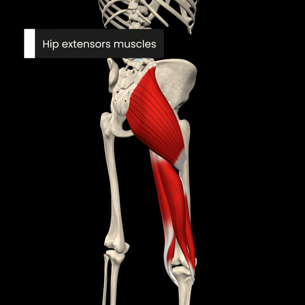
The primary extensor of the hip joint is the gluteus maximus muscle , assisted by the hamstring muscles ( biceps femoris , semitendinosus , semimembranosus ) and the adductor magnus muscle .
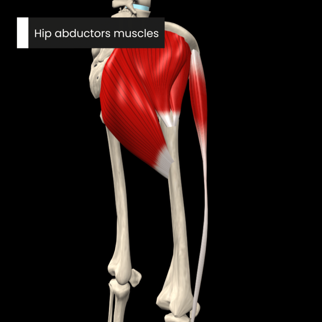
The primary abductors of the hip joint are the gluteus medius and the gluteus minimus muscles. The tensor fasciae latae, piriformis and sartorius muscles also assist in hip abduction. The hip abductors play an active role in stabilizing the pelvis during specific phases of the gait cycle .
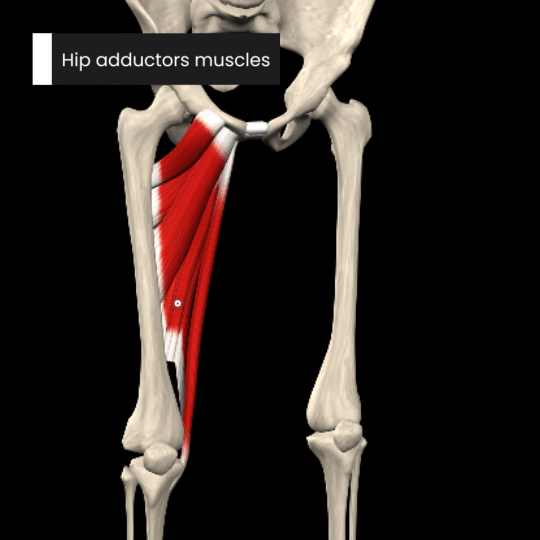
The major adductors of the hip joint are the adductors longus, brevis and magnus and the gracilis muscle . These are assisted by pectineus, quadratus femoris and the inferior fibres of gluteus maximus.
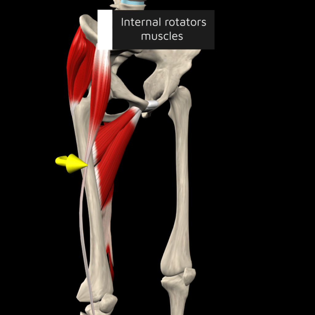
The anterior fibres of glutei minimus and medius are the principal muscles responsible for internal rotation of the hip joint. These muscles are assisted by the tensor fasciae latae and most adductor muscles.
External rotation is produced by the gluteus maximus together with a group of 6 small muscles (lateral rotators): piriformis, obturator internus , superior and inferior gemelli , quadratus femoris and obturator externus .
Learn more about the hip joint by exploring our articles, video tutorials, quizzes and labelled diagrams from this study unit.
:format(jpeg)/images/study_unit/anatomy-hip-joint/QIDFi4AcDDh5LRKoMon86Q_HipJoint_Thumbnail_05.png)
References:
- Magee, D. J. (2014). Orthopedic physical assessment (6th ed.). St. Louis: Elsevier Saunders.
- McKinley, M. & Loughlin, V. (2012). Human anatomy. New York: McGraw-Hill.
- Moore, K. L., Dalley, A. F., & Agur, A. M. R. (2014). Clinically Oriented Anatomy (7th ed.). Philadelphia, PA: Lippincott Williams & Wilkins.
- Netter, F. (2019). Atlas of Human Anatomy (7th ed.). Philadelphia, PA: Saunders.
- Palastanga, N., & Soames, R. (2012). Anatomy and human movement: structure and function (6th ed.). Edinburgh: Churchill Livingstone.
- Standring, S. (2016). Gray's Anatomy (41st ed.). Edinburgh: Elsevier Churchill Livingstone.
Illustrators:
- Hip joint - Liene Znotina
Hip joint : want to learn more about it?
Our engaging videos, interactive quizzes, in-depth articles and HD atlas are here to get you top results faster.
What do you prefer to learn with?
“I would honestly say that Kenhub cut my study time in half.” – Read more.

Learning anatomy isn't impossible. We're here to help.
Learning anatomy is a massive undertaking, and we're here to help you pass with flying colours.
Want access to this video?
- Curated learning paths created by our anatomy experts
- 1000s of high quality anatomy illustrations and articles
- Free 60 minute trial of Kenhub Premium!
...it takes less than 60 seconds!
Want access to this quiz?
Want access to this gallery.
The Hip Joint
Written by Oliver Jones
Last updated January 21, 2022 • 41 Revisions •
The hip joint is a ball and socket synovial joint, formed by an articulation between the pelvic acetabulum and the head of the femur.
It forms a connection from the lower limb to the pelvic girdle, and thus is designed for stability and weight-bearing – rather than a large range of movement.
In this article, we shall look at the anatomy of the hip joint – its articulating surfaces, ligaments and neurovascular supply.
Premium Feature
Structures of the hip joint, articulating surfaces.
The hip joint consists of an articulation between the head of femur and acetabulum of the pelvis.
The acetabulum is a cup-like depression located on the inferolateral aspect of the pelvis. Its cavity is deepened by the presence of a fibrocartilaginous collar – the acetabular labrum . The head of femur is hemispherical, and fits completely into the concavity of the acetabulum.
Both the acetabulum and head of femur are covered in articular cartilage, which is thicker at the places of weight bearing.
The capsule of the hip joint attaches to the edge of the acetabulum proximally. Distally, it attaches to the intertrochanteric line anteriorly and the femoral neck posteriorly.
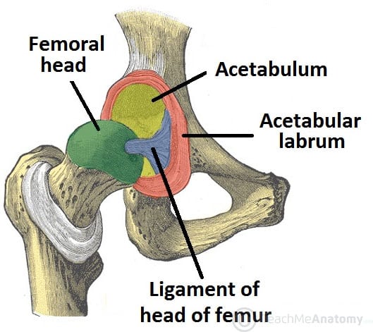
Fig 1 The articulating surfaces of the hip joint – pelvic acetabulum and head of the femur.
The ligaments of the hip joint act to increase stability. They can be divided into two groups – intracapsular and extracapsular:
Intracapsular
The only intracapsular ligament is the ligament of head of femur . It is a relatively small structure, which runs from the acetabular fossa to the fovea of the femur.
It encloses a branch of the obturator artery (artery to head of femur), a minor source of arterial supply to the hip joint.
Extracapsular
There are three main extracapsular ligaments, continuous with the outer surface of the hip joint capsule:
- It has a ‘Y’ shaped appearance, and prevents hyperextension of the hip joint. It is the strongest of the three ligaments.
- It has a triangular shape, and prevents excessive abduction and extension.
- It has a spiral orientation, and prevents hyperextension and holds the femoral head in the acetabulum.
Neurovascular Supply
The arterial supply to the hip joint is largely via the medial and lateral circumflex femoral arteries – branches of the profunda femoris artery (deep femoral artery). They anastomose at the base of the femoral neck to form a ring, from which smaller arteries arise to supply the hip joint itself.
The medial circumflex femoral artery is responsible for the majority of the arterial supply (the lateral circumflex femoral artery has to penetrate through the thick iliofemoral ligament). Damage to the medial circumflex femoral artery can result in avascular necrosis of the femoral head.
The artery to head of femur and the superior/inferior gluteal arteries provide some additional supply.
The hip joint is innervated primarily by the sciatic, femoral and obturator nerves. These same nerves innervate the knee, which explains why pain can be referred to the knee from the hip and vice versa.
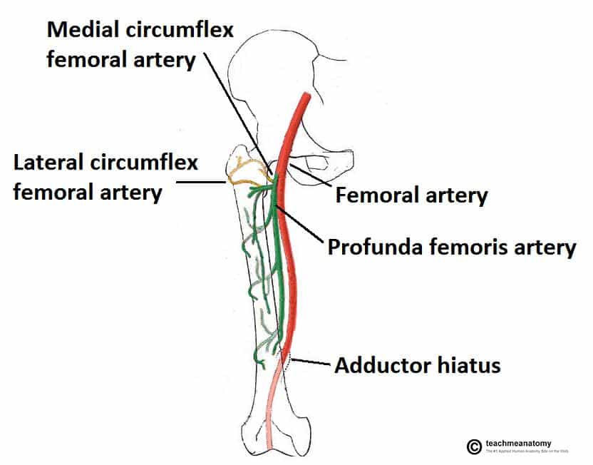
Fig 2 The medial and lateral circumflex femoral arteries are the major blood supply to the hip joint.
Stabilising Factors
The primary function of the hip joint is to weight-bear . There are a number of factors that act to increase stability of the joint.
The first structure is the acetabulum . It is deep, and encompasses nearly all of the head of the femur. This decreases the probability of the head slipping out of the acetabulum (dislocation).
There is a horseshoe shaped fibrocartilaginous ring around the acetabulum which increases its depth, known as the acetabular labrum . The increase in depth provides a larger articular surface, further improving the stability of the joint.
The iliofemoral, pubofemoral and ischiofemoral ligaments are very strong, and along with the thickened joint capsule, provide a large degree of stability. These ligaments have a unique spiral orientation ; this causes them to become tighter when the joint is extended.
In addition, the muscles and ligaments work in a reciprocal fashion at the hip joint:
- Anteriorly , where the ligaments are strongest, the medial flexors (located anteriorly) are fewer and weaker.
- Posteriorly , where the ligaments are weakest, the medial rotators are greater in number and stronger – they effectively ‘pull’ the head of the femur into the acetabulum.
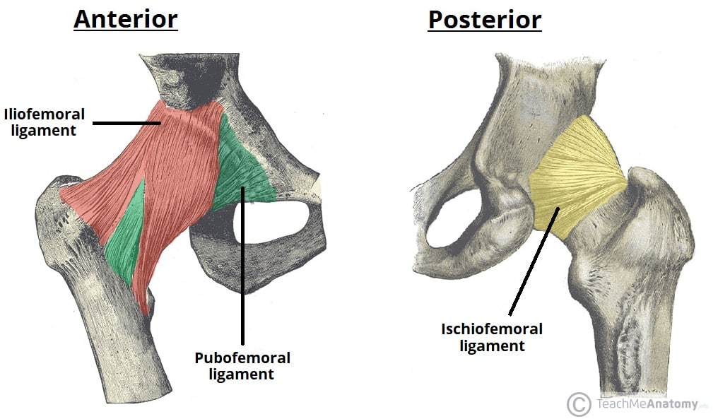
Fig 3 The extracapsular ligaments of the hip joint; ileofemoral, pubofemoral and ischiofemoral ligaments.
Movements and Muscles
The movements that can be carried out at the hip joint are listed below, along with the principle muscles responsible for each action:
- Flexion – iliopsoas, rectus femoris, sartorius, pectineus
- Extension – gluteus maximus; semimembranosus, semitendinosus and biceps femoris (the hamstrings)
- Abduction – gluteus medius, gluteus minimus, piriformis and tensor fascia latae
- Adduction – adductors longus, brevis and magnus, pectineus and gracilis
- Lateral rotation – biceps femoris, gluteus maximus, piriformis, assisted by the obturators, gemilli and quadratus femoris.
- Medial rotation – anterior fibres of gluteus medius and minimus, tensor fascia latae
The degree to which flexion at the hip can occur depends on whether the knee is flexed – this relaxes the hamstring muscles , and increases the range of flexion.
Extension at the hip joint is limited by the joint capsule and the iliofemoral ligament . These structures become taut during extension to limit further movement.
Clinical Relevance
Dislocation of the hip joint, congenital dislocation.
Congenital hip dislocation occurs as a result of developmental dysplasia of the hip (DDH). It occurs when the Acetabulum is shallow as a result of failure to develop properly in utero
Common clinical features include:
- Limited abduction at the hip joint
- Limb length discrepancy – the affected limb is shorter
- Asymmetrical gluteal or thigh skin folds
DDH is usually treated with a Pavlik harness . This holds the femoral head in the acetabular fossa and promotes normal development of the hip joint. Surgery is indicated in cases that do not respond to harness treatment.
Acquired Dislocation
Acquired dislocations of the hip joint are relatively uncommon, owing to the strength and stability of the joint. They usually occur as a result of trauma , but it can occur as a complication following Total Hip Replacement or hemiarthroplasty.
There are two main types of acquired hip dislocation; posterior and anterior:
- The affected limb becomes shortened and medially rotated.
- The sciatic nerve runs posteriorly to the hip joint, and is at risk of injury (occurs in 10-20% of cases). This is often associated with anterior femoral head and posterior wall fractures
- Anterior dislocation (rare) – occurs as a consequence of traumatic extension, abduction and lateral rotation. The femoral head is displaced anteriorly and (usually) inferiorly in relation to the acetabulum.
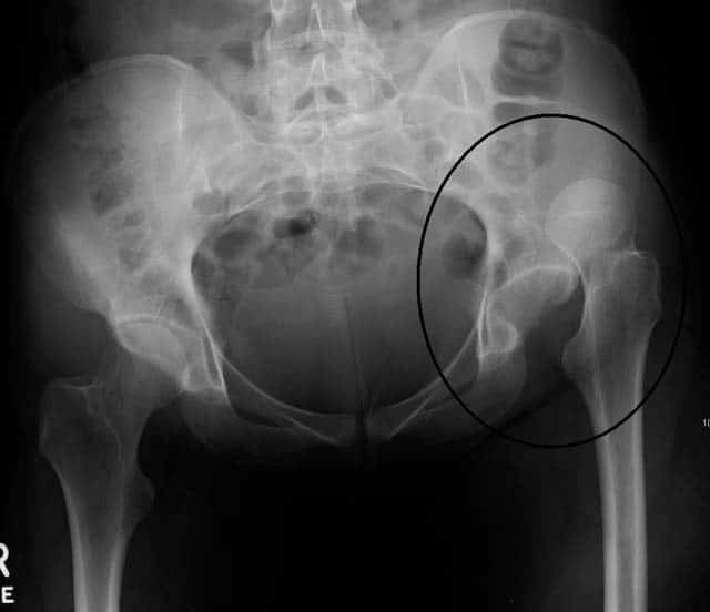
Fig 4 Radiograph showing dislocation of the left hip joint.
Do you think you’re ready? Take the quiz below
Question 1 of 3
Recommended Reading
Forgot password.
Please enter your username or email address below. You will receive a link to create a new password via emai and please check that the email hasn't been delivered into your spam folder.
We use cookies to improve your experience on our site and to show you relevant advertising. To find out more, read our privacy policy .
Privacy Overview
Report question, rate this article.
- Anatomy & Physiology
Home » Blog » Anatomy & Physiology » Anatomy of the Hip Joint: Bones, Ligaments, and Muscles
Anatomy of the Hip Joint: Bones, Ligaments, and Muscles
- May 30, 2024
- 6 minutes read
The hip joint is fundamental to human movement, enabling us to walk, run, and carry out countless daily activities. As the most proximal of the lower extremity joints, the hip plays a vital role in weight-bearing and provides a wide range of motion. This detailed Muscle and Motion article will explore the hip joint’s anatomy and functions and how it plays an integral role in human movement.
Anatomy of the hip joint
The hip is a ball-and-socket joint where the rounded head of the femur fits snugly into the acetabulum of the pelvis. This structure allows multiple movements, including flexion, extension, abduction, adduction, and internal and external rotation.
Ligaments and capsule structure
The hip joint is surrounded by a strong fibrous capsule that forms a sleeve around the acetabulum and the neck of the femur. The key ligaments that provide stability are:
- Iliofemoral Ligament : Shaped like a Y, this ligament is crucial for maintaining an upright posture by limiting hyperextension. It is particularly important for standing and supporting individuals with spinal cord injuries.
- Pubofemoral Ligament: Stretching across the joint medially and inferiorly, this ligament limits hyperextension and abduction.
- Ischiofemoral Ligament: Located on the posterior aspect of the joint, this ligament prevents excessive internal rotation and adduction when the hip is flexed.
These ligaments are strategically attached in a spiral, allowing them to be flexible during leg flexion and tight during extension. This mechanism enables standing without muscle effort, instead relying on the iliofemoral ligament, which is especially beneficial for individuals with paralysis from spinal cord injuries.
Acetabular labrum
The acetabular labrum is a fibrocartilaginous structure that surrounds the rim of the acetabulum, forming a ring around the socket of the hip joint. This ring significantly deepens the socket, increasing its concavity and creating a more secure and stable fit for the femoral head. Functionally, the labrum is vital in maintaining the joint’s overall stability by preventing dislocation and enhancing the distribution of joint forces. It also plays a crucial role in sustaining intra-articular pressure, helping to seal the hip joint and retain the lubricating synovial fluid. The labrum’s unique structure also absorbs shock, protecting the joint from damage.
Hip joint stability and positioning
- Close-Packed Position : The hip joint achieves its highest stability when close-packed, characterized by the hip being fully extended, adducted, and internally rotated. In this state, the hip ligaments are taut, and the joint’s bony anatomy aligns to provide maximal stability, securely holding the femoral head within the acetabulum. This positioning creates tension in the ligaments and is crucial for weight-bearing activities like standing upright. This position also limits excessive movement, protecting the joint from dislocations and injuries.
- Open-Packed Position: The open-packed position occurs during hip flexion, abduction, and external rotation. This position, known as the “hook-lying” position, relaxes the hip ligaments, reducing the compressive forces on the joint and increasing the joint capsule’s laxity. However, while offering improved flexibility, the open-packed position sacrifices some joint stability due to the loose ligamentous and capsular structures.
Femoral angles and their impact
Two critical angles in the femur play a substantial role in determining hip joint function and lower limb alignment: the angle of inclination and the angle of torsion.
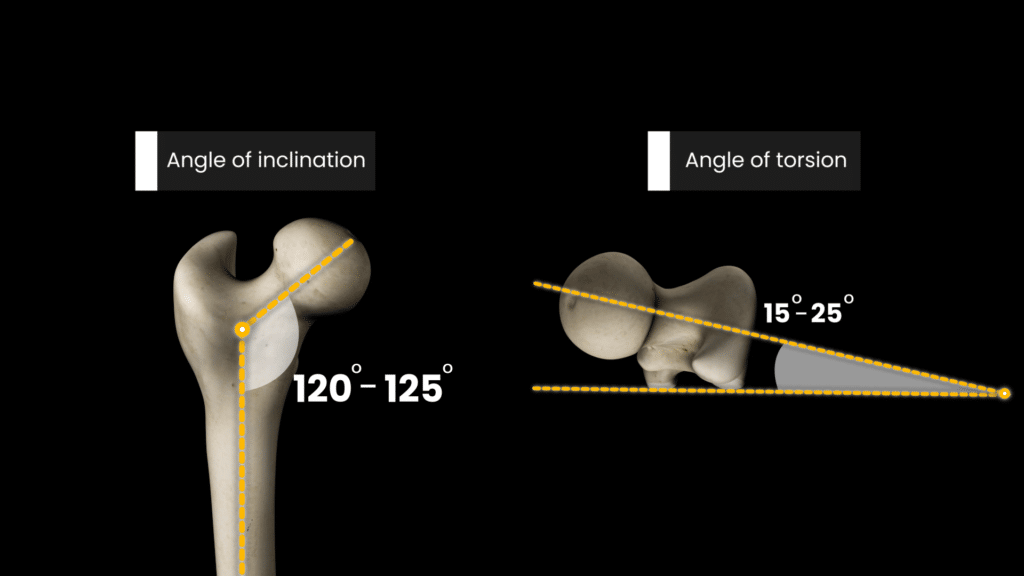
Angle of inclination
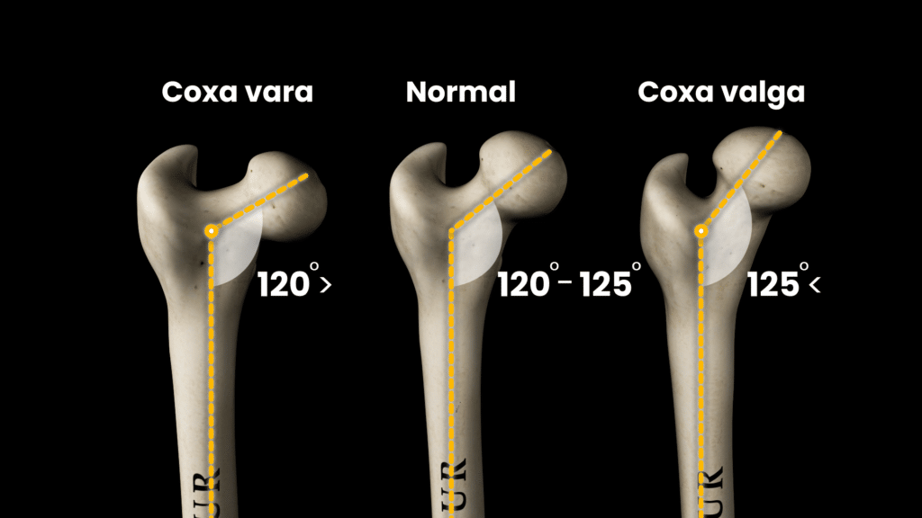
The angle of inclination, created between the femoral neck and the femoral shaft, generally ranges from 120 to 125 degrees in adults. This angle is essential because it influences how the femoral head sits within the acetabulum and how weight is distributed across the hip joint.
- Coxa Valga : An angle above 125 degrees, leading to a straighter femoral neck.
- Coxa Vara: An angle below 120 degrees, resulting in a more horizontal femoral neck
Angle of torsion
The femoral angle of torsion is the outward twist of the femoral head and neck from the shaft and typically ranges between 15 to 25 degrees. When viewed from above, the femoral head and neck align over the shaft, marked by a line through the distal femoral condyles. This angle is crucial as it dictates the hip’s rotation and, consequently, the walking pattern.
- Anteversion: An increase in this angle results in inward-facing hips, often causing a “toed-in” gait.
- Retroversio n : Decreased angle leads to outward-facing hips and a “toed-out” gait.
Hip joint range of motion
Understanding the normal ranges of motion for the hip joint is crucial for assessing joint health and function. Here’s a detailed look at the typical active range of motion for the hip:
- Flexion – 120 degrees
- Extension – 10 degrees
- Abduction – 45 degrees
- Adduction – 25 degrees
- Internal rotation – 15 degrees
- External rotation – 35 degrees
These values represent the active range of motion, meaning they describe how far the joint can move by muscle action alone without any external assistance.
Muscle function and movement
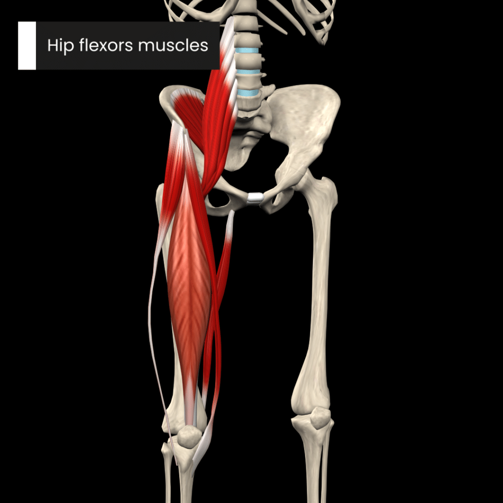
The hip muscles play a pivotal role in generating and controlling the movements of the joint:
Hip flexor muscles
- Psoas major
- Psoas minor
- Rectus femoris
- Tensor fasciae latae
Hip extensors muscles
- Gluteus maximus
Hip adductors muscles
- Adductor magnus
- Adductor longus
- Adductor brevis
Hip abductors muscles
- Gluteus medius
- Gluteus minimus
Hip internal rotator muscles
- Adductor magnus (hamstring part)
Hip external rotator muscles
- Gemellus superior
- Gemellus inferior
- Obturator externus
- Obturator internus
- Quadratus femoris
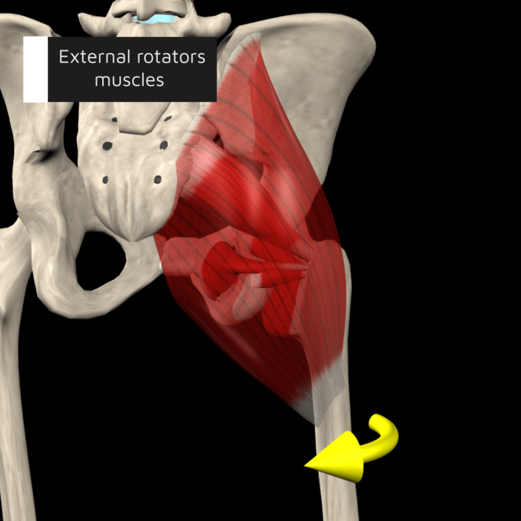
In summary, the hip joint is an anatomical marvel that plays a crucial role in human movement and weight-bearing activities. Its ball-and-socket structure allows for a remarkable range of motion while the surrounding ligaments, muscles, and the acetabular labrum provide stability and protection. The arrangement of the joint capsule and ligaments enables secure weight distribution and mobility.
Check out our comprehensive Strength Training App (all anatomy content included in the strength app) to learn more about the fascinating world of hip anatomy and how each muscle contributes to movement. Our app provides detailed visualizations and interactive models to help you understand the complex interactions of hip muscles, ligaments, and bones.
Whether you’re a student eager to master anatomy, a professional looking to refresh your knowledge, or simply curious about how your body moves, our app offers a wealth of information at your fingertips.
Explore at your own pace, learn through engaging content, and discover the intricate mechanics of the hip joint that support your daily activities. Visit our website to access the app and start your journey into the dynamic world of human anatomy today!
Dive deeper into our content – Download our Strength Training App Today.
All anatomy content is included in the Strength Training App.
- Glenister, R., & Sharma, S. (2023). Anatomy, bony pelvis and lower limb, hip. StatPearls Publishing.
- Netter. (2018). Atlas of Human Anatomy (7th ed.). Elsevier – Health Sciences Division.
- Lim, W. (2021). Clinical application and limitations of the capsular pattern. Physical Therapy Korea, 28(1), 13–17.
- Privacy Overview
- Strictly Necessary Cookies
This website uses cookies so that we can provide you with the best user experience possible. Cookie information is stored in your browser and performs functions such as recognising you when you return to our website and helping our team to understand which sections of the website you find most interesting and useful.
Strictly Necessary Cookie should be enabled at all times so that we can save your preferences for cookie settings.
If you disable this cookie, we will not be able to save your preferences. This means that every time you visit this website you will need to enable or disable cookies again.
eOrthopod.com
…clear, understandable information about muscles, bones and joints
Hip Anatomy
A patient’s guide to hip anatomy, introduction.
The hip joint is a true ball-and-socket joint. This arrangement gives the hip a large amount of motion needed for daily activities like walking, squatting, and stair-climbing.
Understanding how the different layers of the hip are built and connected can help you understand how the hip works, how it can be injured, and how challenging recovery can be when this joint is injured. The deepest layer of the hip includes the bones and the joints. The next layer is made up of the ligaments of the joint capsule. The tendons and the muscles come next.
In addition to reading this article, be sure to watch our Hip Anatomy Animated Tutorial Video .
This guide will help you understand
- the parts that make up the hip
- how these parts work together
Important Structures
The important structures of the hip can be divided into several categories. These include
- bones and joints
- ligaments and tendons
- blood vessels
Bones and Joints
The bones of the hip are the femur (the thighbone) and the pelvis . The top end of the femur is shaped like a ball. This ball is called the femoral head . The femoral head fits into a round socket on the side of the pelvis. This socket is called the acetabulum .
The femoral head is attached to the rest of the femur by a short section of bone called the femoral neck . A large bump juts outward from the top of the femur, next to the femoral neck. This bump, called the greater trochanter , can be felt along the side of your hip. Large and important muscles connect to the greater trochanter. One muscle is the gluteus medius . It is a key muscle for keeping the pelvis level as you walk.
Articular cartilage is the material that covers the ends of the bones of any joint. Articular cartilage is about one-quarter of an inch thick in the large, weight-bearing joints like the hip. Articular cartilage is white and shiny and has a rubbery consistency. It is slippery, which allows the joint surfaces to slide against one another without causing any damage. The function of articular cartilage is to absorb shock and provide an extremely smooth surface to make motion easier. We have articular cartilage essentially everywhere that two bony surfaces move against one another, or articulate .
In the hip, articular cartilage covers the end of the femur and the socket portion of the acetabulum in the pelvis. The cartilage is especially thick in the back part of the socket, as this is where most of the force occurs during walking and running.
Ligaments and Tendons
There are several important ligaments in the hip. Ligaments are soft tissue structures that connect bones to bones. A joint capsule is a watertight sac that surrounds a joint. In the hip, the joint capsule is formed by a group of three strong ligaments that connect the femoral head to the acetabulum. These ligaments are the main source of stability for the hip. They help hold the hip in place.
A small ligament connects the very tip of the femoral head to the acetabulum. This ligament, called the ligamentum teres , doesn’t play a role in controlling hip movement like the main hip ligaments. It does, however, have a small artery within the ligament that brings a very small blood supply to part of the femoral head.
A long tendon band runs alongside the femur from the hip to the knee. This is the iliotibial band . It gives a connecting point for several hip muscles. A tight iliotibial band can cause hip and knee problems.
A special type of ligament forms a unique structure inside the hip called the labrum . The labrum is attached almost completely around the edge of the acetabulum. The shape and the way the labrum is attached create a deeper cup for the acetabulum socket. This small rim of cartilage can be injured and cause pain and clicking in the hip.
The hip is surrounded by thick muscles. The gluteals make up the muscles of the buttocks on the back of the hip. The inner thigh is formed by the adductor muscles . The main action of the adductors is to pull the leg inward toward the other leg. The muscles that flex the hip are in front of the hip joint. These include the iliopsoas muscle . This deep muscle begins in the low back and pelvis and connects on the inside edge of the upper femur. Another large hip flexor is the rectus femoris . The rectus femoris is one of the quadriceps muscles, the largest group of muscles on the front of the thigh. Smaller muscles going from the pelvis to the hip help to stabilize and rotate the hip.
Finally, the hamstring muscles that run down the back of the thigh start on the bottom of the pelvis. Because the hamstrings cross the back of the hip joint on their way to the knee, they help to extend the hip, pulling it backwards.
All of the nerves that travel down the thigh pass by the hip. The main nerves are the femoral nerve in front and the sciatic nerve in back of the hip. A smaller nerve, called the obturator nerve , also goes to the hip.
These nerves carry the signals from the brain to the muscles that move the hip. The nerves also carry signals back to the brain about sensations such as touch, pain, and temperature.
Blood Vessels
Traveling along with the nerves are the large vessels that supply the lower limb with blood. The large femoral artery begins deep within the pelvis. It passes by the front of the hip area and goes down toward the inner edge of the knee. If you place your hand on the front of your upper thigh you may be able to feel the pulsing of this large artery.
The femoral artery has a deep branch, called the profunda femoris ( profunda means deep). The profunda femoris sends two vessels that go through the hip joint capsule. These vessels are the main blood supply for the femoral head. As mentioned earlier, the ligamentum teres contains a small blood vessel that gives a very small supply of blood to the top of the femoral head.
Other small vessels form within the pelvis and supply the back portion of the buttocks and hip.
Where friction occurs between muscles, tendons, and bones there is usually a structure called a bursa . A bursa is a thin sac of tissue that contains fluid to lubricate the area and reduce friction. The bursa is a normal structure. The body will even produce a bursa in response to friction.
Think of a bursa like this. If you press your hands together and slide them against one another, you produce some friction. In fact, when your hands are cold you may rub them together briskly to create heat from the friction. Now imagine that you hold in your hands a small plastic sack that contains a few drops of salad oil. This sack would let your hands glide freely against each other without a lot of friction.
A bursa that sometimes causes problems in the hip is sandwiched between the bump on the outer hip (the greater trochanter) and the muscles and tendons that cross over the bump. This bursa, called the greater trochanteric bursa , can get irritated if the iliotibial band (discussed earlier) is tight. Another bursa sits between the iliopsoas muscle where it passes in front of the hip joint. Bursitis here is called iliopsoas bursitis . A third bursa is over the ischial tuberosity , the bump of bone in your buttocks that you sit on.>
As you can see, the hip is complex with a design that provides a good amount of stability. It allows good mobility and range of motion for doing a wide range of daily activities. Many powerful muscles connect to and cross by the hip joint, making it possible for us to accelerate quickly during actions like running and jumping.
134k followers
13k followers
290k followers
839k followers
Hip Assessment

- Extremity Online Course
The Hip Joint
The hip joint is a deep ball and socket joint comprised of the convex femoral head and the concave acetabulum of the pelvis. Similar to the glenohumeral joint, the acetabulum has a glenoidal lip or labrum around its edges for extra stability.

Epidemiology
It is estimated that around 10% of the general population suffers from some sort of hip complaint ( Birrel et al. 2005 ). In the athletic population (mainly club football) hip/groin injuries accounted for 4-19% of all injuries ( Weir et al. 2015 ). The most common conditions, which we will cover in more detail in this course are osteoarthritis, labral tears due to femoroacetabular impingement (FAI), and greater trochanteric pain syndrome (GTPS) also known as gluteal tendinopathy.
According to the Dutch study by Picavet et al. (2003) , around 28% of people with hip or knee pain have continuous mild pain and mild pain is recurrent in 46% of people with either a hip or knee complaint.
Lievense et al. (2007) followed 224 patients age ≥50 with hip pain over a period of six years and assessed disease progression and incidence of total hip replacement at three and six years. After three years, 15% of patients showed disease progression and 12% underwent hip replacement surgery. Disease progression almost doubled at the second follow-up (28%) and 22% had received a hip replacement.
The individual prognosis for the specific pathologies is discussed in the next units.
Prognostic factors
According to a review by Artus et al. (2017) the following factors are related, but not limited to, the course of hip pain:
- widespread pain
- high functional disability
- somatization
- high pain intensity
- presence of previous pain episodes
Next to the general red flags that should be asked in any case, specific red flags in the hip joint can be:
1) Region-specific red flags:
- Again fractures. This is especially important in the elderly population where hip fractures accounted for up to 14% of all fractures ( Burge et al. 2007 , Ensrud et al. 2013 )
- Avascular femoral head necrosis, which manifests as intermittent groin pain that increases with activity. Usually the case in prolonged use of corticosteroids ( Barille et al. 2014 )
2) Tract anamnesis:
- Gastrointestinal tract: this visceral pain is mostly deep aching, gnawing, vague, or deep grinding. Conditions include GI bleeding, epigastric pain, constipation, obturator or psoas abscess, appendicitis.
Furthermore, you should not forget to rule out SI Joint or lumbar spine involvement in case of a hip complaint. This is because the hip receives innervation from spinal segments L2-S2. Therefore it is a common site for referred pain from these structures. Check the “further reading” section and the bottom for literature on the topic.
LEVEL UP YOUR DIFFERENTIAL DIAGNOSIS IN RUNNING RELATED HIP PAIN – FOR FREE!
Basic Assessment
Depending on the outcome, your basic assessment can give you the following info: 1) Limitations in range of motion and their end-feel can guide structural assessment (e.g. bone to bone=osteoarthritis, empty=tendinopathy due to pain)
It’s best to start with Active Range of Motion Assessment:
Standard values for the range of motion in different directions are as follows:

AROM assessment is then typically followed by Passive Range of Motion Assessment (PROM). PROM assessment in the hip joint is also used as part of the diagnostic criteria for hip osteoarthritis, where a marked decrease increases the likelihood of OA.
During PROM assessment, it’s important to compare the range of motion as well as the end-feel of the affected hip with the unaffected side.
Muscle strains or sprains are common in the hip and groin region, especially in athletic populations. In the following two videos, you will see how you can stress the muscles around the hip joint to possibly identify them as a source of nociception as well as a video on the differentiation of groin pain in athletes.

Specific Pathologies in the Hip
There are several pathologies that are commonly seen in the hip area. For more information, click on the respective pathology (content will be added in the near future):
- Muscle Contractures
- Leg Length Difference
- Hip Microinstability
- Femoroacetabular Impingement (FAI) / Labrum Tears
- Tendinopathy
- Deep Gluteal Syndrome
- Ischiofemoral Impingement
- Osteoarthritis of the Hip
Artus, Majid et al. “Generic Prognostic Factors for Musculoskeletal Pain in Primary Care: A Systematic Review.” BMJ Open 7.1 (2017): e012901. PMC . Web. 6 Sept. 2018.
Birrell F, Croft P, Cooper C, Hosie G, Macfarlane GJ & Silman A. (2000) Radiographic change is common in new presenters in primary care with hip pain. Rheumatology. 2000;39:772–5.
Burge R, Dawson-Hughes B, Solomon DH, Wong JB, King A & Tosteson A. (2007) Incidence and economic burden of osteoporosis-related fractures in the United States, 2005-2025. J Bone Miner Res. 2007;22:465–475.
Barille MF, Wu JS & McMahon CJ. (2014) Femoral head avascular necrosis: a frequently missed incidental finding on multidetector CT. Clin Radiol. 2014 Mar;69(3):280–5.
Ensrud KE. (2013) Epidemiology of fracture risk with advanced age. Gerontol A Biol Sci Med Sci 2013;68(10):1236–1242
Lievense, Annet M., et al. “Prognosis of hip pain in general practice: a prospective followup study.” Arthritis Care & Research 57.8 (2007): 1368-1374.
Picavet, H. S. J., and J. S. A. G. Schouten. “Musculoskeletal pain in the Netherlands: prevalences, consequences and risk groups, the DMC3-study.” Pain 102.1-2 (2003): 167-178.
Weir, Adam, et al. “Doha agreement meeting on terminology and definitions in groin pain in athletes.” Br J Sports Med 49.12 (2015): 768-774.
Follow a course
- Learn from wherever, whenever, and at your own pace
- Interactive online courses from an award-winning team
- CEU/CPD accreditation in the Netherlands, Belgium, US & UK
How to Boost Your Knowledge about the 23 Most Common Orthopedic Pathologies in Just 40 Hours

What customers have to say about this online course

Create your free account to gain access to this exclusive content and more!
To provide the best experiences, we and our partners use technologies like cookies to store and/or access device information. Consenting to these technologies will allow us and our partners to process personal data such as browsing behavior or unique IDs on this site and show (non-) personalized ads. Not consenting or withdrawing consent, may adversely affect certain features and functions.
Click below to consent to the above or make granular choices. Your choices will be applied to this site only. You can change your settings at any time, including withdrawing your consent, by using the toggles on the Cookie Policy, or by clicking on the manage consent button at the bottom of the screen.
Download our free physiotherapy app with all the knowledge you need.


Please Enable JavaScript in your Browser to Visit this Site.
We are there to help you, click on any available representative to contact 🌸

DaBaby Takes Plea Deal to Avoid Jail Time in 'Sucker Punch' Case

Kendrick Lamar 'Not Like Us' Video Earns Rapper Free Tam's Burgers For Life

Run-DMC's Darryl McDaniels Wants Alternative to Trump vs. Biden

Common, Pete Rock Perform with The Roots, Posdnuos, Bilal on 'Tonight Show'

Diddy's Mother Janice Hospitalized with Chest Pains
Kendrick lamar 'not like us' boosts tam's burgers' business 40 percent, kendrick lamar beef selling beef 'not like us' boosts compton 🍔 joint's sales.
Kendrick Lamar highlighting his hometown burger joint in his "Not Like Us" video is paying big dividends ... as the folks at Tam's Burgers say they're stacking bread and cheese, just like their signature dish!
TB manager Lauro Hernandez and his son Bryan Noe tell TMZ Hip Hop ... customers have been flooding their Rosecrans Ave location in Compton all weekend, after K. Dot released the smash music vid on July 4.
They say many fans who came in, told them "Not Like Us" was the reason for their visit -- and while they still have steady support from locals, Lauro and Bryan say they're picking up lots of tourist business.
Business is up 30% to 40% since the video dropped .
In the video, Kendrick and 'NLU' producer Mustard pull up to Tam's in a Lambo and post up inside with dancer Storm DeBarge during the song's 2nd verse.
Waiting for your permission to load the Instagram Media.
Lauro says people have been largely ordering the bacon cheeseburger -- that’s what Kendrick gets when he's there.
He's known Kendrick since he was a teenager, having been grounded in the community for the past 20 years, and he finds it amazing the superstar rapper comes back and never forgets his roots.
Kendrick's shouted out Tam's over the years in his songs, even on his most recent "Mr. Morale & the Big Steppers" album, and Lauro tells us he happily shut down for the day when Dot and Dave Free came to film scenes inside.
They didn’t ask for any money for using the restaurant in the video, but Kendrick wanted to make sure they got something for their time, and for closing down -- so, they agreed the Tam's logo would be featured in the final cut.
That look's clearly paying off, based on the sales boost at Tam's -- which adds up when you see the music video already has more than 35 million views! That's a ton of free advertising.
Kendrick's homies also wanted to take care of him ... Jason Martin and Jay Worthy went half on billboards in Compton featuring the city-unifying images from June's Pop Out concert at the Forum.
The West Coast is having a modern-day renaissance!!!
- Share on Facebook
related articles

Recording Academy CEO Harvey Mason Jr. Says 'Not Like Us' Diss Eligible For Grammys

Jason Martin Recounts Kendrick Lamar 'Not Like Us' Video, Bobbi Althoff Cameo
Old news is old news be first.
- Manage Account
Kendrick Helps Burger Joint Featured in ‘Not Like Us’ Video Boost Sales
Tam's Burgers No. 21 was swamped when we called them up to confirm.
By Angel Diaz
- Share on Facebook
- Share to Flipboard
- Share on Pinterest
- + additional share options added
- Share on Reddit
- Share on LinkedIn
- Share on Whats App
- Send an Email
- Print this article
- Post a Comment
- Share on Tumblr
The Kendrick stimmy pack is the gift that keeps on giving.
Tam’s Burgers No. 21 — located on Rosecrans Ave. in his hometown of Compton — has seen a major uptick in business after being prominently featured in his video for “ Not Like Us ,” according to TMZ . We called and confirmed that they’ve seen around a 40 percent increase in sales with most customers ordering Kendrick’s go-to bacon cheeseburger. They also told us they’re thinking about naming a menu item after the Compton rapper.
Joe Jonas Asked For Jonas Brothers' 'Blessings' to Make Upcoming Solo Album
Kendrick lamar.
See latest videos, charts and news
Trending on Billboard
We can’t give Kendrick all the credit here, though. New Ho King was featured prominently in the video of Drake’s response “ Family Matters .” The restaurant’s owner can be seen wearing Pharrell’s jewelry that the Toronto rapper notoriously bought in auction.
Tam’s was also featured on the set of Dr. Dre and Snoop Dogg’s Super Bowl 56 Halftime Show.
Get weekly rundowns straight to your inbox
Want to know what everyone in the music business is talking about?
Get in the know on.
Billboard is a part of Penske Media Corporation. © 2024 Billboard Media, LLC. All Rights Reserved.
optional screen reader
Charts expand charts menu.
- Billboard Hot 100™
- Billboard 200™
- Hits Of The World™
- TikTok Billboard Top 50
- Songs Of The Summer
- Song Breaker
- Year-End Charts
- Decade-End Charts
Music Expand music menu
- R&B/Hip-Hop
Videos Expand videos menu
Culture expand culture menu, media expand media menu, business expand business menu.
- Business News
- Record Labels
- View All Pro
Pro Tools Expand pro-tools menu
- Songwriters & Producers
- Artist Index
- Royalty Calculator
- Market Watch
- Industry Events Calendar
Billboard Español Expand billboard-espanol menu
- Cultura y Entretenimiento
Get Up Anthems by Tres Expand get-up-anthems-by-tres menu
Honda music expand honda-music menu.

Health | Robotic technology allows Mercy Health surgeons…
Share this:.
- Click to share on Facebook (Opens in new window)
- Click to share on X (Opens in new window)
- Lorain County
- Cuyahoga County
- Opioid Epidemic
Health | Robotic technology allows Mercy Health surgeons to personalize joint replacement procedures

The new equipment is located at Mercy Health — Allen Hospital, attached to Oberlin Primary Care, and will transform the way total knee, partial knee and total hip replacements are performed, by helping surgeons know more and cut less, aiding in precision, recovery time and improved outcomes, the release said.
Mako SmartRobotics combines three key components: 3D CT-based planning; AccuStopTM haptic technology; and insightful data analytics into one platform that has shown better outcomes for total knee, total hip and partial knee patients, according to the release.
“With Mako SmartRobotics, I know more about my patients than ever before, and I’m able to cut less,” said Mercy Health orthopedic surgeon Dr. Joseph George, in the release. “For some patients, this can mean less soft tissue damage; for others, greater bone preservation.
“Mako’s 3D CT allows me to create a personalized plan based on each patient’s unique anatomy before entering the operating room. During surgery, I can validate that plan and make any necessary adjustments while guiding the robotic arm to execute that plan.
“It’s exciting to be able to offer this transformative technology across the joint replacement service line to perform total knee, and total hip replacements.”
Total knee replacements in the United States are expected to increase 189% by 2030, yet studies have shown that approximately 20% of patients are dissatisfied after conventional surgery, according to the release.
Mako Total Knee combines Stryker’s advanced robotic technology with its clinically successful Triathlon Total Knee System, which enables surgeons to have a more predictable surgical experience with increased precision and accuracy, the release said.
In clinical studies, Mako Total Knee demonstrated the potential for patients to experience less pain, less need for opiate analgesics, less need for inpatient physical therapy, reduction in length of hospital stay, improved knee flexion and greater soft tissue protection in comparison to manual techniques, according to the release.
By 2030, total hip replacements in the United States are projected to grow 171%. Mako SmartRobotics for Total Hip is a treatment option for adults who suffer from degenerative joint disease of the hip, the release said.
During surgery, the surgeon guides the robotic arm during bone preparation to prepare the hip socket and position the implant according to the predetermined surgical plan.
In a controlled matched-paired study to measure acetabular bone resection, results suggested greater bone preservation for Mako Total Hip compared to manual surgery, the release said.
“We are proud to offer this highly advanced SmartRobotics technology in our area,” George said. “This addition to our orthopedic service line further demonstrates our commitment to provide the community with outstanding health care.”
To learn more about Mercy Health’s orthopedic services, visit mercy.com .
More in Health

Health | Lack of affordability tops older Americans’ list of health care worries

Health | Beyond PMS: A poorly understood disorder means periods of despair for some women

Health | Patient sings along to rock song during first awake kidney transplant at Northwestern Medicine

Health | Mounjaro bests Ozempic for weight loss in first head-to-head comparison of real-world use

COMMENTS
Hip joint (Articulatio coxae) The hip joint is a ball and socket type of synovial joint that connects the pelvic girdle to the lower limb. In this joint, the head of the femur articulates with the acetabulum of the pelvic (hip) bone.. The hip joint is a multiaxial joint and permits a wide range of motion; flexion, extension, abduction, adduction, external rotation, internal rotation and ...
Study with Quizlet and memorize flashcards containing terms like Proximal hamstring release., Partial hip replacement with bipolar arthroplasty to treat right hip fracture., Removal of a foreign body deep in the left thigh. and more.
Study with Quizlet and memorize flashcards containing terms like Push up, Squat, Dead lift and more.
The hip joint is a ball and socket synovial joint, formed by an articulation between the pelvic acetabulum and the head of the femur.. It forms a connection from the lower limb to the pelvic girdle, and thus is designed for stability and weight-bearing - rather than a large range of movement.. In this article, we shall look at the anatomy of the hip joint - its articulating surfaces ...
Piriformis. In summary, the hip joint is an anatomical marvel that plays a crucial role in human movement and weight-bearing activities. Its ball-and-socket structure allows for a remarkable range of motion while the surrounding ligaments, muscles, and the acetabular labrum provide stability and protection. The arrangement of the joint capsule ...
Ligaments, Blood supply, Relations, Nerve supply, Movements, Muscles associated with movements and Clinical anatomy of the Hip joint.To get the notes of anat...
A Patient's Guide to Hip Anatomy Introduction The hip joint is a true ball-and-socket joint. This arrangement gives the hip a large amount of motion needed for daily activities like walking, squatting, and stair-climbing. Understanding how the different layers of the hip are built and connected can help you understand how the hip works, how
Hip Joint Week 11. The hip joint is one of the most important joints in the human body, and it is responsible for facilitating movement in the lower extremities. The hip joint is a ball-and-socket joint, with the head of the femur (thigh bone) fitting into a socket in the pelvis known as the acetabulum.
KINES 3092 Assignment 6: Hip joint exercise movement analysis -After analyzing each of the exercises in the hip joint movement analysis chart, break each into two primary movement phases such as a lifting phase and lowering phase.-For each phase, determine which hip joint movements occur, and then list the hip joint muscles responsible for causing those movements.
PROM assessment in the hip joint is also used as part of the diagnostic criteria for hip osteoarthritis, where a marked decrease increases the likelihood of OA. During PROM assessment, it's important to compare the range of motion as well as the end-feel of the affected hip with the unaffected side. Muscle strains or sprains are common in the ...
This assignment will be graded on knowledge and understanding (30%), thinking Assessment Chart for Joint Overlay, Sports Medicine or Joint Construction Assignment Level 1 Level 2 Level 3 Level 4 Knowledge and Understanding Instructions are not fully understood or followed Determines little of the criteria by which the success of the
THE HIP JOINT Muscles of the Hip Gluteus Maximus O: lower posterior iliac crest and posterior surface of the sacrum I: gluteal tuberosity (upper, posterior aspect of the femur) & I.T. band Actions: Extension of the hip External rotation of the hip Lower fibers (below the center of motion) assist in adduction Gluteus Maximus Produces hip extension beyond 15 degrees; not used extensively during ...
Week 8: Chapter 8 Homework/Assignment. While standing, twisting your lower limb at the hip so that your toes point off to the side is called _______ rotation. Twisting your lower limb at the hip so that your toes point toward your other foot is called _______ rotation. A.) medial; lateral. B.) lateral; medial.
Hip and Knee Joint Movement Assignment. University: Life University (GA) Course: Student (32017) 10 Documents. Info More info. Download. Save. 5a111e4f ce85954ac281c3366f379a51.doc x. Instructions: 1) Find your name in th e list f or y our assigned mov ement. 2) Pr epare the scrip t f or your Fl ipGrid® video.
Perform any technique of your choice showed in the hip joint chapters given above and record a video of yourself doing the technique. The video should not be more than 40-60 seconds. So, prepare your model/patient and your position before starting the recording. Make sure the angle of recording is appropriate for instructor to understand […]
Running head: Written Assignment Unit 2 (Jones, 2019). The hip joint is a ball-and-socket synovial type of joint, wherein the ball is the femoral head of the femur and the socket, which is acetabulum of the pelvis (Jones, 2019). It is the articulation of the pelvis with the femur where the axial skeleton is connected with the lower extremity. Adult coxae or hip bone is fusio of three (3) bones ...
Assignment (hip joint) - Read online for free.
Assignment 3 - Hip joint Yuanyuan Cai T00539672 Part 1: factors contributing to the stability and mobility of the hip joint, the same as you have done for the shoulder joint on assignment 2. Part 2: comparing shoulder and hip joints regarding all the factors of stability and mobility.-Hip joint: Hip joint is a ball and socket synovial joint where the head of femur attach to the acetabulum of ...
CH. 13: Exercise 13.7. Proximal hamstring release. For code 27097, go to CPT index main term Release, subterm Muscle, and qualifier Hamstring. Verify the code in the Repair, Revision, and/or Reconstruction subcategory of the Pelvis and Hip Joint category in the Musculoskeletal subsection of the Surgery section.
Kendrick Lamar highlighting his hometown burger joint in his "Not Like Us" video is paying big dividends ... as the folks at Tam's Burgers say they're stacking bread and cheese, just like their ...
R&B/Hip-Hop; 07/10/2024 ... In the video, Dot and Mustard pull up to the local burger joint in a black 2021 Ferrari SF90 Stradale for a quick break during the song's second verse.
View Muscle Assignment 3.docx from ANAT 35A at Mt San Antonio College. 2.1.4 The hip joint and ligaments around it Name the ligaments that form the hip joint capsule. Ilio-femoral and ischio-femoral
By 2030, total hip replacements in the United States are projected to grow 171%. Mako SmartRobotics for Total Hip is a treatment option for adults who suffer from degenerative joint disease of the ...